Each mammary complex consists of 5-20 mammary units and their corresponding ducts. …
Each mammary complex consists of 5-20 mammary units and their corresponding ducts. The ducts open separately on the tip of the teat. Shallow grooves indicate the border between complexes. An intermammary sulcus divides the right from the left row.
Active transport is reliant on carrier proteins and thus follows the same …
Active transport is reliant on carrier proteins and thus follows the same rules as facilitated diffusion in that they are specific have a maximum rate and are subject to competition. Crucially they transport substances against their concentration gradient and so require energy to work.
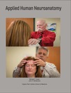
The course will start with an overview of the central and peripheral …
The course will start with an overview of the central and peripheral nervous systems (CNS and PNS), the development of their structure and major divisions. The major functional components of the CNS will then be reviewed individually. Topography, functional distribution of nerve cell bodies, ascending and descending tracts in the spinal cord. Brainstem organization and functional components, including cranial nerve nuclei, ascending / descending pathways, amine-containing cells, structure and information flow in the cerebellar and vestibular systems. Distribution of the cranial nerves, resolution of their skeletal and branchial arch components. Functional divisions of the Diencephalon and Telencephalon. The course will then continue with how these various CNS pieces and parts work together. Motor systems, motor neurons and motor units, medial and lateral pathways, cortical versus cerebellar systems and their functional integration. The sensory systems, visual, auditory and somatosensory. Olfaction will be covered in the context of the limbic system, which will also include autonomic control and the Papez circuit. To conclude, functional organization and information flow in the neocortex will be discussed.
Mechanical forces play a decisive role during development of tissues and organs, …
Mechanical forces play a decisive role during development of tissues and organs, during remodeling following injury as well as in normal function. A stress field influences cell function primarily through deformation of the extracellular matrix to which cells are attached. Deformed cells express different biosynthetic activity relative to undeformed cells. The unit cell process paradigm combined with topics in connective tissue mechanics form the basis for discussions of several topics from cell biology, physiology, and medicine.
This resource is a video abstract of a research paper created by …
This resource is a video abstract of a research paper created by Research Square on behalf of its authors. It provides a synopsis that's easy to understand, and can be used to introduce the topics it covers to students, researchers, and the general public. The video's transcript is also provided in full, with a portion provided below for preview:
"Our genome is a lot like a book. When cells need information critical to their function, they must physically crack the genome open to arrive at the right chapter, or gene sequence. Often, the relevance of details from chapter one isn’t clear until chapter ten. Similarly, non-coding sequences often control the expression of genes far away in linear genomic distance but relatively close in three-dimensional space. Now, a new method of probing these regions could help scientists gain more information from much less starting material—and, in the process, help us learn more about the book of life. The technique is called HiCAR, short for Hi-C on Accessible Regulatory DNA. HiCAR builds off the Hi-C method, which uses high-throughput sequencing to detect how different regions of genomic DNA interact with each other. Specifically, HiCAR targets the regions of chromatin that are open and accessible to proteins with information about gene regulation..."
The rest of the transcript, along with a link to the research itself, is available on the resource itself.
This resource is a video abstract of a research paper created by …
This resource is a video abstract of a research paper created by Research Square on behalf of its authors. It provides a synopsis that's easy to understand, and can be used to introduce the topics it covers to students, researchers, and the general public. The video's transcript is also provided in full, with a portion provided below for preview:
"Physicians may soon be able to get a detailed look at blood vessels surrounding breast tumors quickly, painlessly, and without radiation, thanks to the work of a team of Japanese researchers. One application of the technology is earlier and more accurate tracking of when cancer has turned deadly. The formation of new blood vessels around a tumor is a key sign that cancer is getting ready to spread. But getting a clear look at these blood vessels can be tricky. Approaches like MRI or computed tomography often come with a hefty price tag, and exposure to contrast agent or radiation may pose health risks. To sidestep these issues, the researchers optimized a way to perform photoacoustic imaging. This type of imaging utilizes the light-absorbing properties of hemoglobin to show where blood is flowing in the body. When hemoglobin is exposed to pulses of laser light, it produces small vibrations. These vibrations are picked up by scanners and used to generate a detailed map of blood vessel architecture..."
The rest of the transcript, along with a link to the research itself, is available on the resource itself.
This resource is a video abstract of a research paper created by …
This resource is a video abstract of a research paper created by Research Square on behalf of its authors. It provides a synopsis that's easy to understand, and can be used to introduce the topics it covers to students, researchers, and the general public. The video's transcript is also provided in full, with a portion provided below for preview:
"A new report published in Arthritis & Rheumatology suggests that cardiovascular disease affects patients with systemic lupus erythematosus much earlier than previously thought – in some cases even before active lupus sets in. The finding was reported by a team of researchers based in China who have been working on validating the use of magnetic resonance imaging to detect the early manifestations of cardiac impairment. With heart disease being the leading cause of death in patients with SLE, the ability to detect very early signs of cardiac dysfunction in this group could one day lay a foundation for enhanced preventive strategies. Traditional cardiac MRI approaches like late gadolinium enhancement don’t perform well in detecting early indications of heart disease, such as disturbances in myocardial extracellular volume. Missing these early warning signs could hide the fact that heart disease has set in and potentially complicate treatment efforts..."
The rest of the transcript, along with a link to the research itself, is available on the resource itself.
This resource is a video abstract of a research paper created by …
This resource is a video abstract of a research paper created by Research Square on behalf of its authors. It provides a synopsis that's easy to understand, and can be used to introduce the topics it covers to students, researchers, and the general public. The video's transcript is also provided in full, with a portion provided below for preview:
"Light-responsive proteins have revolutionized our understanding of the brain. By introducing the genes encoding these proteins into neurons and then exciting the cells using lasers – a technique known as optogenetics – individual cells can be rapidly turned on or off, enabling exquisitely sensitive investigations of brain function. But a fundamental limitation of the method is that light doesn’t travel very far through brain tissue, which has hampered the study of more buried – and often vital – structures. Now, researchers at the RIKEN Center for Brain Science have developed a way to extend the reach of optogenetics by nearly an order of magnitude, providing new possibilities for deep-brain stimulation. The team accomplished this using a special type of nanoparticle known as an upconversion nanoparticle, so named for its ability to transform – or “upconvert” – near-infrared light into visible output..."
The rest of the transcript, along with a link to the research itself, is available on the resource itself.
This resource is a video abstract of a research paper created by …
This resource is a video abstract of a research paper created by Research Square on behalf of its authors. It provides a synopsis that's easy to understand, and can be used to introduce the topics it covers to students, researchers, and the general public. The video's transcript is also provided in full, with a portion provided below for preview:
"Increased blood flow to the uterus during pregnancy is essential to the health of both mother and baby But the cellular processes that promote blood flow during pregnancy aren’t fully understood Now, researchers have discovered that fat surrounding blood vessels in the uterus plays a key role In pregnant rats, uterine blood flow was up to 3 times higher than in non-pregnant rats But blood flow plummeted when fat tissue was removed from the uterus of pregnant rats Interestingly, tests on isolated vessels demonstrated that fat tissue-shrinking factors could be at play which seems counterintuitive because narrow vessels generally mean low blood flow One explanation is that isolating tissue from its natural surroundings could produce changes not observed in a live animal Future studies will explore this apparent contradiction and hopefully reveal the role of fat tissue in human pregnancy Osikoya et al..."
The rest of the transcript, along with a link to the research itself, is available on the resource itself.
This resource is a video abstract of a research paper created by …
This resource is a video abstract of a research paper created by Research Square on behalf of its authors. It provides a synopsis that's easy to understand, and can be used to introduce the topics it covers to students, researchers, and the general public. The video's transcript is also provided in full, with a portion provided below for preview:
"This tiny brain structure is known as the claustrum. For more than a hundred years, scientists have speculated about what exactly the claustrum does. But only recently has state-of-the-art biological technology allowed researchers to probe its anatomy and connections to the rest of the brain. Francis Crick—of DNA fame—and neuroscientist Christof Koch hypothesized the claustrum to be the seat of consciousness, a conductor of sorts, orchestrating the activity of neurons in charge of higher brain functions from deep within. Now, new research from the RIKEN Center for Brain Science in Japan appears to confirm that hypothesis. Only, instead of arousing neurons to action, the claustrum lulls them to sleep. The claustrum is both an appropriate and unfortunate name for this important part of the brain’s anatomy. Latin for “hidden or shut away,” the claustrum has long defied close examination due to its thin, irregular shape and placement deep within the brain..."
The rest of the transcript, along with a link to the research itself, is available on the resource itself.
The most recent knowledge of the anatomy, physiology, biochemistry, biophysics, and bioengineering …
The most recent knowledge of the anatomy, physiology, biochemistry, biophysics, and bioengineering of the gastrointestinal tract and the associated pancreatic, liver and biliary tract systems is presented and discussed. Gross and microscopic pathology and the clinical aspects of important gastroenterological diseases are then presented, with emphasis on integrating the molecular, cellular and pathophysiological aspects of the disease processes to their related symptoms and signs.
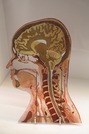
The American Association of Anatomy (AAA) has put forth learning objectives for …
The American Association of Anatomy (AAA) has put forth learning objectives for the four preclinical courses in the anatomical sciences. In conjunction, they have also provided a list of clinical syndromes and scenarios that medical students should understand during their preclinical studies. This resource uses these clinical syndromes as a guide to provide students with a quick reference to clinical syndromes covered in preclinical neuroanatomy. This is part one of three, addressing issues related to gross brain, embryology, and spinal cord functions.This project supported by the Touro OER Faculty Fellowship. Created by Stephanie Klinesmith, Department of Anatomy, Touro College of Osteopathic Medicine - Middletown Campus, 60 Prospect Ave, Middletown, NY, 10940. sklinesm@touro.edu
The American Association of Anatomy (AAA) has put forth learning objectives for …
The American Association of Anatomy (AAA) has put forth learning objectives for the four preclinical courses in the anatomical sciences. In conjunction, they have also provided a list of clinical syndromes and scenarios that medical students should understand during their preclinical studies. This resource uses these clinical syndromes as a guide to provide students with a quick reference to clinical syndromes covered in preclinical neuroanatomy. This is part two of three, addressing issues related to the brainstem and cranial nerves.This project supported by the Touro OER Faculty Fellowship. Created by Stephanie Klinesmith, Department of Anatomy, Touro College of Osteopathic Medicine - Middletown Campus, 60 Prospect Ave, Middletown, NY, 10940. sklinesm@touro.edu
This 155-page manual is comprised of two types of learning activities: 1) …
This 155-page manual is comprised of two types of learning activities: 1) free response fill-in-the blank questions focused of the facts and principles of neuroanatomy and neurophysiology that underpin the neurologic examination and specifically developed exercises that demonstrate how the facts and principles are related to the particular tests and procedures that comprise the neurologic examination. Free response questions form the bulk of the Neuroscience Review section of each chapter and are intended as a review of information previously or concurrently being learned regarding the structure, function and organization of the nervous system. Some questions focus on anatomical or physiological facts and relationships that help explain why certain techniques are performed as they are, such as why non-nociceptive tactile stimuli are required in order to activate nerve impulse transmission in the lemniscal system. Other questions are intended to revisit facts and concepts that are needed to properly interpret the elicited findings. 2) The Application Exercises of each chapter are designed to demonstrate how neuroanatomical and neurophysiological information is used in the design of particular clinical tests of neurologic function. The application exercises are also intended to help users learn how to perform and become comfortable with the various clinical maneuvers and tests that comprise the routine neurologic examination. An important outcome of performing these exercises is that, as a member of a learning group, each individual has the opportunity to experience the neurologic examination from the point of view of the subject (patient)—an experience that arguably provides insight and understanding that can be gained in no other way.
Teeth develop differently in different regions of the mouth in most species, …
Teeth develop differently in different regions of the mouth in most species, a process called heterodonty. In some animals, teeth develop identically in different regions of the mouth, a process called homodonty. Different species will have varying numbers of teeth and different shapes depending largely on their diet. Not all species possess teeth and there is huge variation in dental formulae between the species that have teeth. Teeth are mainly used for mastication - chewing and grinding food particles, but are also used for seizing prey and tearing. The occlusion surface is where opposing teeth touch. The contact surface is where adjacent teeth touch.
Thermoregulation is the ability of an endothermic organism to maintain a relatively …
Thermoregulation is the ability of an endothermic organism to maintain a relatively constant body temperature, despite fluctuations in temperature of the external environment. This is a vital part of homeostasis.
The ileum is the terminal portion of the small intestine and continues …
The ileum is the terminal portion of the small intestine and continues from the jejunum. It opens into the caecum at the ileocaecal orifice. The intestinal epithelium is mainly absorptive, with much less digestion occurring compared to the duodenum and the jejunum.
The rabbit is a monogastric hindgut fermenter and is a herbivore. Microbes …
The rabbit is a monogastric hindgut fermenter and is a herbivore. Microbes in the hindgut produce volatile fatty acids (VFAs) which are available to the animal for energy. Microbes also produce vitamins and protein, which are available only in minimal quantities as they are produced in the hindgut (see advantages and disadvantages of hid gut fermentation). Most microbial fermentation occurs in the caecum (as opposed to the horse where most occurs in the colon). Rabbits usually eat at dusk.
No restrictions on your remixing, redistributing, or making derivative works. Give credit to the author, as required.
Your remixing, redistributing, or making derivatives works comes with some restrictions, including how it is shared.
Your redistributing comes with some restrictions. Do not remix or make derivative works.
Most restrictive license type. Prohibits most uses, sharing, and any changes.
Copyrighted materials, available under Fair Use and the TEACH Act for US-based educators, or other custom arrangements. Go to the resource provider to see their individual restrictions.