Beginning kindergarteners are introduced to science and engineering concepts through questions such …
Beginning kindergarteners are introduced to science and engineering concepts through questions such as “What is a Scientist?” and “What is an Engineer?”, and go on to compare and contrast the two. They are introduced to five steps of the engineering design process and explore these steps using the “I do, we do, you do” set of guided instruction. At the end of the project, students produce a set of purple popsicles that they design using various materials and by following a set of criteria.
Biology is designed for multi-semester biology courses for science majors. It is …
Biology is designed for multi-semester biology courses for science majors. It is grounded on an evolutionary basis and includes exciting features that highlight careers in the biological sciences and everyday applications of the concepts at hand. To meet the needs of today’s instructors and students, some content has been strategically condensed while maintaining the overall scope and coverage of traditional texts for this course. Instructors can customize the book, adapting it to the approach that works best in their classroom. Biology also includes an innovative art program that incorporates critical thinking and clicker questions to help students understand—and apply—key concepts.
By the end of this section, you will be able to:Describe the …
By the end of this section, you will be able to:Describe the role of cells in organismsCompare and contrast light microscopy and electron microscopySummarize cell theory
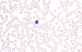
This video shows scanning a Wright's stained blood smear slide with pauses …
This video shows scanning a Wright's stained blood smear slide with pauses to view leukocytes. The video was taken at 630X under a brightfield microscope. This video is compatible with a laboratory lesson in which students observe, categorize, and count leukocytes. More than 100 leukocytes are viewed in this video. Note, this video does not have narration.Video credit: Emily Fox
Use your cell phone to explore the mini-scopic world. Open your eyes …
Use your cell phone to explore the mini-scopic world. Open your eyes to the amazing world of the ultra-tiny when you convert your cell phone into a portable, picture-taking Miniscope using a simple plastic lens from a laser pointer.
In this 7th grade science lesson, students deepen their understanding of pollen …
In this 7th grade science lesson, students deepen their understanding of pollen and pollinators by practicing the use of microscopes to observe pollen and bee species from the garden.
Students are introduced to the latest imaging methods used to visualize molecular …
Students are introduced to the latest imaging methods used to visualize molecular structures and the method of electrophoresis that is used to identify and compare genetic code (DNA). Students should already have basic knowledge of genetics, DNA (DNA structure, nucleotide bases), proteins and enzymes. The lesson begins with a discussion to motivate the need for imaging techniques and DNA analysis, which prepares students to participate in the associated two-part activity: 1) students each choose an imaging method to research (from a provided list of molecular imaging methods), 2) they research basic information about electrophoresis.
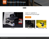
Multi-part open educational resource which includes a video about how to operate …
Multi-part open educational resource which includes a video about how to operate a compound microscope, an interactive walk-through of the parts of a microscope, with detailed descriptions, and a self assessment. In addition to these wonderful instructional tools, there is a virtual lab, the "Lettuce Onion Lab" available on the same page.
This module provides a short introduction to light microscopy using two divergent yeast …
This module provides a short introduction to light microscopy using two divergent yeast species, Saccharomyces cerevisiae and Schizosaccharomyces pombe, as subjects. Students examine the yeasts at different phases of their life cycle and compare them to the much smaller bacterium, E. coli. At the end of the module,students should be able to:identify the components of the light microscopestain samples with iodine to improve contrastcorrectly adjust the microscope for different specimensidentify morphological differences between yeast species in both stationary and vegetative phasesThis module is part of a semester-long introductory laboratory course, Investigations in Molecular Cell Biology, at Boston College.
This micrograph was taken at 1000X total magnifcation on a brightfield microscope. …
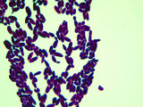
This micrograph was taken at 1000X total magnifcation on a brightfield microscope. The subject is Bacillus cereus cells were grown in broth culture for 48 hours at 30 degrees Celsius. The cells were heat-fixed to a slide and stained with malachite green (endospores) and safranin red (vegetative cells) prior to visualization.Image credit: Emily Fox
This micrograph was taken at 1000X total magnifcation on a brightfield microscope. …
This micrograph was taken at 1000X total magnifcation on a brightfield microscope. The subject is Bacillus megaterium cells were grown in broth culture for 5 days at 30 degrees Celsius. The cells were heat-fixed to a slide and stained with malachite green (endospores) and safranin red (vegetative cells) prior to visualization.Image credit: Emily Fox
This micrograph was taken at 1000X total magnifcation on a brightfield microscope. …
This micrograph was taken at 1000X total magnifcation on a brightfield microscope. The subject is Bacillus subtilis cells were grown in broth culture for 48 hours at 30 degrees Celsius. The cells were heat-fixed to a slide and stained with malachite green (endospores) and safranin red (vegetative cells) prior to visualization.Image credit: Emily Fox
This micrograph was taken at 1000X total magnifcation on a brightfield microscope. …
This micrograph was taken at 1000X total magnifcation on a brightfield microscope. The subject is Bacillus subtilis cells were grown in broth culture for 5 days at 30 degrees Celsius. The cells were heat-fixed to a slide and stained with malachite green (endospores) and safranin red (vegetative cells) prior to visualization.Image credit: Emily Fox
This micrograph was taken at 1000X total magnifcation on a brightfield microscope. …
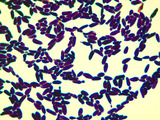
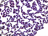
This micrograph was taken at 1000X total magnifcation on a brightfield microscope. The subject is Bacillus subtilis cells grown in broth culture overnight at 30 degrees Celsius. The cells were heat-fixed to a slide Gram stained prior to visualization.Image credit: Emily Fox
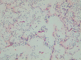
This micrograph was taken at 100X total magnifcation on a brightfield microscope. …
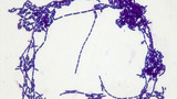
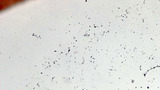
This micrograph was taken at 100X total magnifcation on a brightfield microscope. The subject is Bacillus subtilis cells grown in broth culture overnight at 30 degrees Celsius. The cells were heat-fixed to a slide Gram stained prior to visualization.Image credit: Emily Fox
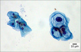
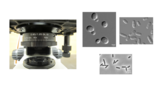
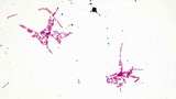
This micrograph was taken at 1000X total magnifcation on a brightfield microscope. …
This micrograph was taken at 1000X total magnifcation on a brightfield microscope. The subject is Candida albicans cells grown in broth culture at 30 degrees Celsius. The cells were heat-fixed to a slide and Gram stained prior to visualization.Image credit: Emily Fox
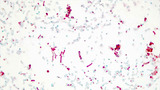
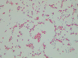
This micrograph was taken at 1000X total magnifcation on a brightfield microscope. …
This micrograph was taken at 1000X total magnifcation on a brightfield microscope. The subject is Candida albicans cells grown on YPD agar at 30 degrees Celsius. The cells were heat-fixed to a slide and Gram stained prior to visualization.Image credit: Emily Fox
No restrictions on your remixing, redistributing, or making derivative works. Give credit to the author, as required.
Your remixing, redistributing, or making derivatives works comes with some restrictions, including how it is shared.
Your redistributing comes with some restrictions. Do not remix or make derivative works.
Most restrictive license type. Prohibits most uses, sharing, and any changes.
Copyrighted materials, available under Fair Use and the TEACH Act for US-based educators, or other custom arrangements. Go to the resource provider to see their individual restrictions.