Biology is designed for multi-semester biology courses for science majors. It is …
Biology is designed for multi-semester biology courses for science majors. It is grounded on an evolutionary basis and includes exciting features that highlight careers in the biological sciences and everyday applications of the concepts at hand. To meet the needs of today’s instructors and students, some content has been strategically condensed while maintaining the overall scope and coverage of traditional texts for this course. Instructors can customize the book, adapting it to the approach that works best in their classroom. Biology also includes an innovative art program that incorporates critical thinking and clicker questions to help students understand—and apply—key concepts.
By the end of this section, you will be able to:Describe the …
By the end of this section, you will be able to:Describe the role of cells in organismsCompare and contrast light microscopy and electron microscopySummarize cell theory
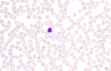
This video shows scanning a Wright's stained blood smear slide with pauses …
This video shows scanning a Wright's stained blood smear slide with pauses to view leukocytes. The video was taken at 630X under a brightfield microscope. This video is compatible with a laboratory lesson in which students observe, categorize, and count leukocytes. More than 100 leukocytes are viewed in this video. Note, this video does not have narration.Video credit: Emily Fox
The purpose of this lesson is to teach students about blood and …
The purpose of this lesson is to teach students about blood and its components while instilling an appreciation of its importance for survival. The lesson takes a step-by-step approach to determining the recipe for blood while introducing students to important laboratory techniques like centrifugation and microscopy, as well as some diseases of cell types found in blood. It also highlights the importance of donating blood by explaining basic physiological concepts and the blood donation procedure.
This resource is a video abstract of a research paper created by …
This resource is a video abstract of a research paper created by Research Square on behalf of its authors. It provides a synopsis that's easy to understand, and can be used to introduce the topics it covers to students, researchers, and the general public. The video's transcript is also provided in full, with a portion provided below for preview:
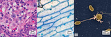
"Our mouths are a vast jungle of microbial life. Here, more than 700 distinct types of microbes make their home, but not everywhere all at once. Each region (the tongue, teeth, gums, etc.) hosts a unique community of microorganisms. To explore this complex living structure, researchers examined the community of bacteria found in the dental plaque of 14 healthy volunteers. Samples indicated the well-known formation of intricate corncob-like structures, where a central filament made of cells of Corynebacteria (magenta) is decorated with “kernels” of spherical Streptococcus bacteria (green). A closer look revealed that these kernels can be composed of a single species of bacteria or contain mixtures of different species. The major corncob species were common to all 14 donors. Corncob composition likely was dictated by the metabolic and binding interactions shared between corncob residents..."
The rest of the transcript, along with a link to the research itself, is available on the resource itself.
Students will learn to fabricate, remix, and design detection and monitoring devices …
Students will learn to fabricate, remix, and design detection and monitoring devices for health following the core focus of the Tricorder: a portable, handheld diagnostic device which can brings health solutions to consumers at home or in remote parts of the world. Inspired by the Tricorder X-Prize (with a purse of $10 million), students will aim to create specific component technologies that integrate into a comprehensive Tricorder mechanism capable of reading vital signs and specific disease biomarker detection. Component areas will include optical, electric, biochemical, and molecular diagnostics.
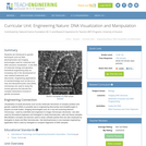
Students are introduced to genetic techniques such as DNA electrophoresis and imaging …
Students are introduced to genetic techniques such as DNA electrophoresis and imaging technologies used for molecular and DNA structure visualization. In the field of molecular biology and genetics, biomedical engineering plays an increasing role in the development of new medical treatments and discoveries. Engineering applications of nanotechnology such as lab-on-a-chip and deoxyribonucleic acid (DNA) microarrays are used to study the human genome and decode the complex interactions involved in genetic processes.
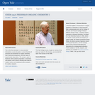
This is the first semester in a two-semester introductory course focused on …
This is the first semester in a two-semester introductory course focused on current theories of structure and mechanism in organic chemistry, their historical development, and their basis in experimental observation. The course is open to freshmen with excellent preparation in chemistry and physics, and it aims to develop both taste for original science and intellectual skills necessary for creative research.
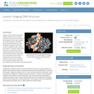
Students are introduced to the latest imaging methods used to visualize molecular …
Students are introduced to the latest imaging methods used to visualize molecular structures and the method of electrophoresis that is used to identify and compare genetic code (DNA). Students should already have basic knowledge of genetics, DNA (DNA structure, nucleotide bases), proteins and enzymes. The lesson begins with a discussion to motivate the need for imaging techniques and DNA analysis, which prepares students to participate in the associated two-part activity: 1) students each choose an imaging method to research (from a provided list of molecular imaging methods), 2) they research basic information about electrophoresis.
This resource is a video abstract of a research paper created by …
This resource is a video abstract of a research paper created by Research Square on behalf of its authors. It provides a synopsis that's easy to understand, and can be used to introduce the topics it covers to students, researchers, and the general public. The video's transcript is also provided in full, with a portion provided below for preview:
"Animals and plants have close relationships with the bacteria on their surfaces and macroalgae — like kelp — are no different. The spatial structure of these microbial communities can impact how they interact with their neighbors, host, and environment. A recent study used spectral imaging to characterize the spatial structure of the bacteria on _Nereocystis luetkeana_. The kelp hosted a dense microbial biofilm that consisted of closely associated, but diverse, microbial taxa. For example, Gammaproteobacteria were found close to the kelp surface, and filamentous Bacteroidetes and Alphaproteobacteria were concentrated near the biofilm-seawater interface. Bacterial density also varied along the length of the kelp blades with density increasing from new tissue at the base to older tissue at the blade tips. Between kelp populations, declining populations hosted fewer microbial cells than kelp from a stable population. This study characterized the dense, spatially differentiated community on _N..."
The rest of the transcript, along with a link to the research itself, is available on the resource itself.
The exercises in this laboratory manual are designed to engage students in …
The exercises in this laboratory manual are designed to engage students in hand-on activities that reinforce their understanding of the microbial world. Topics covered include: staining and microscopy, metabolic testing, physical and chemical control of microorganisms, and immunology. The target audience is primarily students preparing for a career in the health sciences, however many of the topics would be appropriate for a general microbiology course as well.
This resource is a video abstract of a research paper created by …
This resource is a video abstract of a research paper created by Research Square on behalf of its authors. It provides a synopsis that's easy to understand, and can be used to introduce the topics it covers to students, researchers, and the general public. The video's transcript is also provided in full, with a portion provided below for preview:
"During epithelial-mesenchymal transition (EMT), epithelial cells lose their polarity and their cell-cell connections to become mobile, in part via transcription factor (TF) activation. EMT and its reverse process, MET, are critical for tissue development in embryos, and EMT enables wound healing during adulthood, but EMT is also how cancer cells metastasize. Live imaging of animal embryos can yield important insights into these key processes. For example, FGF and actomyosin have been found to regulate intercellular adherens junction (AJ) remodeling during EMT in fruit flies. In addition, in zebrafish embryos, the planar cell polarity (PCP) protein pk1 ensures proper EMT of neural crest cells (NCCs), and cadherin 6 ultimately regulates NCC migration..."
The rest of the transcript, along with a link to the research itself, is available on the resource itself.
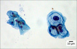
This micrograph was taken at 1000X total magnifcation on a brightfield microscope. …
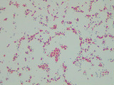
This micrograph was taken at 1000X total magnifcation on a brightfield microscope. The subject is Bacillus cereus cells were grown in broth culture for 48 hours at 30 degrees Celsius. The cells were heat-fixed to a slide and stained with malachite green (endospores) and safranin red (vegetative cells) prior to visualization.Image credit: Emily Fox
This micrograph was taken at 1000X total magnifcation on a brightfield microscope. …
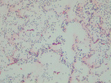
This micrograph was taken at 1000X total magnifcation on a brightfield microscope. The subject is Bacillus megaterium cells were grown in broth culture for 5 days at 30 degrees Celsius. The cells were heat-fixed to a slide and stained with malachite green (endospores) and safranin red (vegetative cells) prior to visualization.Image credit: Emily Fox
This micrograph was taken at 1000X total magnifcation on a brightfield microscope. …
This micrograph was taken at 1000X total magnifcation on a brightfield microscope. The subject is Bacillus subtilis cells were grown in broth culture for 48 hours at 30 degrees Celsius. The cells were heat-fixed to a slide and stained with malachite green (endospores) and safranin red (vegetative cells) prior to visualization.Image credit: Emily Fox
This micrograph was taken at 1000X total magnifcation on a brightfield microscope. …
This micrograph was taken at 1000X total magnifcation on a brightfield microscope. The subject is Bacillus subtilis cells were grown in broth culture for 5 days at 30 degrees Celsius. The cells were heat-fixed to a slide and stained with malachite green (endospores) and safranin red (vegetative cells) prior to visualization.Image credit: Emily Fox
This micrograph was taken at 1000X total magnifcation on a brightfield microscope. …
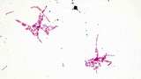
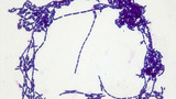
This micrograph was taken at 1000X total magnifcation on a brightfield microscope. The subject is Bacillus subtilis cells grown in broth culture overnight at 30 degrees Celsius. The cells were heat-fixed to a slide Gram stained prior to visualization.Image credit: Emily Fox
This micrograph was taken at 100X total magnifcation on a brightfield microscope. …
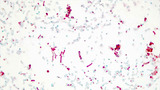
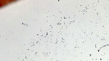
This micrograph was taken at 100X total magnifcation on a brightfield microscope. The subject is Bacillus subtilis cells grown in broth culture overnight at 30 degrees Celsius. The cells were heat-fixed to a slide Gram stained prior to visualization.Image credit: Emily Fox
No restrictions on your remixing, redistributing, or making derivative works. Give credit to the author, as required.
Your remixing, redistributing, or making derivatives works comes with some restrictions, including how it is shared.
Your redistributing comes with some restrictions. Do not remix or make derivative works.
Most restrictive license type. Prohibits most uses, sharing, and any changes.
Copyrighted materials, available under Fair Use and the TEACH Act for US-based educators, or other custom arrangements. Go to the resource provider to see their individual restrictions.