Updating search results...

Search Resources

16 Results

View
Selected filters:
  • medical-imaging
AFNI Training Bootcamp
Conditional Remix & Share Permitted
CC BY-NC-SA
Rating
0.0 stars

This training course is an introduction to the use of the AFNI software suites for the analysis of functional MRI (fMRI) data. It is not intended as an introduction to how fMRI works but is aimed at people who are already doing fMRI data analysis, or those who will be in the near future. 
AFNI (Analysis of Functional NeuroImages) is a leading software suite of C, Python, and R programs and shell scripts, primarily developed for the analysis and display of anatomical and fMRI data. It is freely available for research purposes. 
This event was organized by the Center for Brains, Minds, and Machines (CBMM) Trainee Leadership Council.
CBMM is a multi-institutional NSF Science and Technology Center headquartered at MIT that is dedicated to developing a computationally based understanding of human intelligence and establishing an engineering practice based on that understanding. CBMM brings together computer scientists, cognitive scientists, and neuroscientists to create a new field—the science and engineering of intelligence.

Subject:
Applied Science
Health, Medicine and Nursing
Life Science
Physical Science
Material Type:
Full Course
Provider:
MIT
Provider Set:
MIT OpenCourseWare
Author:
Chen, Gang
Glen, Daniel
Reynolds, Rick
Date Added:
02/01/2018
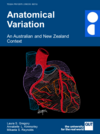
Anatomical Variation: An Australian and New Zealand Context
Conditional Remix & Share Permitted
CC BY-NC
Rating
0.0 stars

Short Description:
This textbook is designed to actively engage your exploration and critical analysis of human anatomical variation in an Australian and New Zealand context. Understanding anatomical variation is essential for all health professionals to avoid patient misdiagnosis such as confusing a natural variant with a pathology, minimise surgical or procedural errors that may occur if variations are unexpected, and ultimately improve patient outcomes by applying culturally safe practices. Research in anatomical variation has demonstrated significant differences in phenotypic expression of variants between and within geographic, ancestral and socioeconomic populations, as well as displaying significant variance between males and females. It is therefore critical as a health professional to understand anatomical variation in the context of the population you intend to practice in. This textbook compiles this critical information into an easy to read summary of the range and frequency of anatomical phenotypes in Australian and New Zealand patients by drawing from contemporary anatomical science research. Anatomical variation of Aboriginal, Torres Strait Islander and Māori peoples has also been highlighted where research is available.

Long Description:
The anatomy of our outwardly facing physical appearance exhibits great diversity between individuals, from different eye, skin and hair colour to the size of our feet and our height. However, it is less known whether our anatomy differs beneath the surface… is the anatomy of the internal organs the same between individuals? Most textbooks would like you to think so with simplified standard descriptions of human anatomy such as the lung lobes and fissures, aortic arch branches and bone numbers. But this eBook is different. Here we build your understanding of the scope and clinical importance of human anatomical variation to improve your clinical skills as a health professional or biomedical scientist.

Anatomical variation is described as the differences in macroscopic morphology (shape and size), topography (location), developmental timing or frequency (number) of an anatomical structure between individuals. It presents during embryological or subadult development and results in no substantive observable interruption to physiological function. Every organ displays an array of anatomical phenotypes, and for these reasons the anatomy of each person is considered a variant. Understanding anatomical variation is essential for all health professionals to avoid patient misdiagnosis such as confusing a natural variant with a pathology, minimise surgical or procedural errors that may occur if variations are unexpected, and ultimately improve patient outcomes by applying culturally safe practices.

This textbook is designed to actively engage your exploration and critical analysis of human anatomical variation in an Australian and New Zealand context. Research in anatomical variation has demonstrated significant differences in phenotypic expression of variants between and within geographic, ancestral and socioeconomic populations, as well as displaying significant variance between males and females. It is therefore critical as a health professional to understand anatomical variation in the context of the population you intend to practice in. This textbook compiles this critical information into an easy to read summary of the range and frequency of anatomical phenotypes in Australian and New Zealand patients by drawing from contemporary anatomical science research. Anatomical variation of Aboriginal, Torres Strait Islander and Māori peoples has also been highlighted where research is available.

The textbook is organised to complement your health science studies by developing your depth of understanding to address three critical themes in anatomical variation: Theme 1: Categorise and describe a range of anatomical variation within the human body. Theme 2: Theorise the implications of anatomical variation on patient outcomes and in professional contexts. Theme 3: Investigate the process of anatomical variation formation and its potential causes.

Each chapter employs a multimodal and active learning approach using text and video summaries of key information, checkpoint quizzes, interactive images, clinical and professional discussion activities, and recommended readings. In this way, the activities in this textbook can be easily embedded into existing health science curricula to strengthen anatomical variation understanding in all health professional courses.

Word Count: 31978

ISBN: 978-1-925553-51-2

(Note: This resource's metadata has been created automatically by reformatting and/or combining the information that the author initially provided as part of a bulk import process.)

Subject:
Anatomy/Physiology
Applied Science
Biology
Education
Health, Medicine and Nursing
Higher Education
Life Science
Material Type:
Textbook
Provider:
Queensland University of Technology
Author:
Annabelle L. Kimmorley
Laura S. Gregory
Mikaela S. Reynolds
Date Added:
06/25/2023
Anatomical Variation: An Australian and New Zealand Context
Conditional Remix & Share Permitted
CC BY-NC
Rating
0.0 stars

Short Description:
This textbook is designed to actively engage your exploration and critical analysis of human anatomical variation in an Australian and New Zealand context. Understanding anatomical variation is essential for all health professionals to avoid patient misdiagnosis such as confusing a natural variant with a pathology, minimise surgical or procedural errors that may occur if variations are unexpected, and ultimately improve patient outcomes by applying culturally safe practices. Research in anatomical variation has demonstrated significant differences in phenotypic expression of variants between and within geographic, ancestral and socioeconomic populations, as well as displaying significant variance between males and females. It is therefore critical as a health professional to understand anatomical variation in the context of the population you intend to practice in. This textbook compiles this critical information into an easy to read summary of the range and frequency of anatomical phenotypes in Australian and New Zealand patients by drawing from contemporary anatomical science research. Anatomical variation of Aboriginal, Torres Strait Islander and Māori peoples has also been highlighted where research is available.

Long Description:
The anatomy of our outwardly facing physical appearance exhibits great diversity between individuals, from different eye, skin and hair colour to the size of our feet and our height. However, it is less known whether our anatomy differs beneath the surface… is the anatomy of the internal organs the same between individuals? Most textbooks would like you to think so with simplified standard descriptions of human anatomy such as the lung lobes and fissures, aortic arch branches and bone numbers. But this eBook is different. Here we build your understanding of the scope and clinical importance of human anatomical variation to improve your clinical skills as a health professional or biomedical scientist.

Anatomical variation is described as the differences in macroscopic morphology (shape and size), topography (location), developmental timing or frequency (number) of an anatomical structure between individuals. It presents during embryological or subadult development and results in no substantive observable interruption to physiological function. Every organ displays an array of anatomical phenotypes, and for these reasons the anatomy of each person is considered a variant. Understanding anatomical variation is essential for all health professionals to avoid patient misdiagnosis such as confusing a natural variant with a pathology, minimise surgical or procedural errors that may occur if variations are unexpected, and ultimately improve patient outcomes by applying culturally safe practices.

This textbook is designed to actively engage your exploration and critical analysis of human anatomical variation in an Australian and New Zealand context. Research in anatomical variation has demonstrated significant differences in phenotypic expression of variants between and within geographic, ancestral and socioeconomic populations, as well as displaying significant variance between males and females. It is therefore critical as a health professional to understand anatomical variation in the context of the population you intend to practice in. This textbook compiles this critical information into an easy to read summary of the range and frequency of anatomical phenotypes in Australian and New Zealand patients by drawing from contemporary anatomical science research. Anatomical variation of Aboriginal, Torres Strait Islander and Māori peoples has also been highlighted where research is available.

The textbook is organised to complement your health science studies by developing your depth of understanding to address three critical themes in anatomical variation: Theme 1: Categorise and describe a range of anatomical variation within the human body. Theme 2: Theorise the implications of anatomical variation on patient outcomes and in professional contexts. Theme 3: Investigate the process of anatomical variation formation and its potential causes.

Each chapter employs a multimodal and active learning approach using text and video summaries of key information, checkpoint quizzes, interactive images, clinical and professional discussion activities, and recommended readings. In this way, the activities in this textbook can be easily embedded into existing health science curricula to strengthen anatomical variation understanding in all health professional courses.

Word Count: 31376

(Note: This resource's metadata has been created automatically by reformatting and/or combining the information that the author initially provided as part of a bulk import process.)
Subject:
Anatomy/Physiology
Applied Science
Biology
Education
Health, Medicine and Nursing
Higher Education
Life Science
Material Type:
Textbook
Provider:
Queensland University of Technology
Date Added:
01/26/2024
Biomedical Information Technology
Conditional Remix & Share Permitted
CC BY-NC-SA
Rating
0.0 stars

This course teaches the design of contemporary information systems for biological and medical data. Examples are chosen from biology and medicine to illustrate complete life cycle information systems, beginning with data acquisition, following to data storage and finally to retrieval and analysis. Design of appropriate databases, client-server strategies, data interchange protocols, and computational modeling architectures. Students are expected to have some familiarity with scientific application software and a basic understanding of at least one contemporary programming language (e.g. C, C++, Java, Lisp, Perl, Python). A major term project is required of all students. This subject is open to motivated seniors having a strong interest in biomedical engineering and information system design with the ability to carry out a significant independent project.
This course was offered as part of the Singapore-MIT Alliance (SMA) program as course number SMA 5304.

Subject:
Applied Science
Biology
Business and Communication
Computer Science
Engineering
Health, Medicine and Nursing
Life Science
Material Type:
Full Course
Provider:
MIT
Provider Set:
MIT OpenCourseWare
Author:
Bhowmick, Sourav
Dewey, C.
Yu, Hanry
Date Added:
09/01/2008
Biomedical Signal and Image Processing
Conditional Remix & Share Permitted
CC BY-NC-SA
Rating
0.0 stars

This course presents the fundamentals of digital signal processing with particular emphasis on problems in biomedical research and clinical medicine. It covers principles and algorithms for processing both deterministic and random signals. Topics include data acquisition, imaging, filtering, coding, feature extraction, and modeling. The focus of the course is a series of labs that provide practical experience in processing physiological data, with examples from cardiology, speech processing, and medical imaging. The labs are done in MATLAB® during weekly lab sessions that take place in an electronic classroom. Lectures cover signal processing topics relevant to the lab exercises, as well as background on the biological signals processed in the labs.

Subject:
Applied Science
Career and Technical Education
Electronic Technology
Engineering
Health, Medicine and Nursing
Material Type:
Full Course
Provider:
MIT
Provider Set:
MIT OpenCourseWare
Author:
Clifford, Gari
Fisher, John
Greenberg, Julie
Wells, William
Date Added:
02/01/2007
Career Options for Biomedical Research
Conditional Remix & Share Permitted
CC BY-NC-SA
Rating
0.0 stars

This course has been designed as a seminar to give students an understanding of how scientists with medical or scientific degrees conduct research in both hospital and academic settings. There will be interactive discussions with research clinicians and scientists about the career opportunities and research challenges in the biomedical field, which an MIT student might prepare for by obtaining an MD, PhD, or combined degrees. The seminar will be held in a case presentation format, with topics chosen from the radiological sciences, including current research in magnetic resonance imaging, positron emission tomography and other nuclear imaging techniques, and advances in radiation therapy. With the lectures as background, we will also examine alternative and related options such as biomedical engineering, medical physics, and medical engineering. We'll use as examples and points of comparisons the curriculum paths available through MIT's Department of Nuclear Science and Engineering. In past years we have given very modest assignments such as readings in advance of or after a seminar, and a short term project.

Subject:
Applied Science
Engineering
Environmental Science
Health, Medicine and Nursing
Material Type:
Full Course
Provider:
MIT
Provider Set:
MIT OpenCourseWare
Author:
He, Xin
Rosen, Bruce
Yip, Sidney
Date Added:
09/01/2006
Functional Magnetic Resonance Imaging: Data Acquisition and Analysis
Conditional Remix & Share Permitted
CC BY-NC-SA
Rating
0.0 stars

This team-taught multidisciplinary course provides information relevant to the conduct and interpretation of human brain mapping studies. It begins with in-depth coverage of the physics of image formation, mechanisms of image contrast, and the physiological basis for image signals. Parenchymal and cerebrovascular neuroanatomy and application of sophisticated structural analysis algorithms for segmentation and registration of functional data are discussed. Additional topics include: fMRI experimental design including block design, event related and exploratory data analysis methods, and building and applying statistical models for fMRI data; and human subject issues including informed consent, institutional review board requirements and safety in the high field environment.
Additional Faculty
Div Bolar
Dr. Bradford Dickerson
Dr. John Gabrieli
Dr. Doug Greve
Dr. Karl Helmer
Dr. Dara Manoach
Dr. Jason Mitchell
Dr. Christopher Moore
Dr. Vitaly Napadow
Dr. Jon Polimeni
Dr. Sonia Pujol
Dr. Bruce Rosen
Dr. Mert Sabuncu
Dr. David Salat
Dr. Robert Savoy
Dr. David Somers
Dr. A. Gregory Sorensen
Dr. Christina Triantafyllou
Dr. Wim Vanduffel
Dr. Mark Vangel
Dr. Lawrence Wald
Dr. Susan Whitfield-Gabrieli
Dr. Anastasia Yendiki

Subject:
Applied Science
Career and Technical Education
Electronic Technology
Engineering
Health, Medicine and Nursing
Material Type:
Full Course
Provider:
MIT
Provider Set:
MIT OpenCourseWare
Author:
Gollub, Randy
Date Added:
09/01/2008
Introduction to Solid-State Chemistry
Conditional Remix & Share Permitted
CC BY-NC-SA
Rating
0.0 stars

In this course, we will explore what makes things in the world the way they are and why, to understand the science and consider the engineering. We learn not only why the physical world behaves the way it does, but also how to think with chemical intuition, which can’t be gained simply by observing the macroscopic world.
This 2018 version of 3.091 by Jeffrey Grossman and the 2010 OCW version by Don Sadoway cover similar topics and both provide complete learning materials. This 2018 version also includes Jeffrey Grossman’s innovative Goodie Bags, Why This Matters, and CHEMATLAS content, as well as additional practice problems, quizzes, and exams.

Subject:
Applied Science
Chemistry
Engineering
Physical Science
Material Type:
Full Course
Provider:
MIT
Provider Set:
MIT OpenCourseWare
Author:
Grossman, Jeffrey
Date Added:
09/01/2018
MEG Workshop
Conditional Remix & Share Permitted
CC BY-NC-SA
Rating
0.0 stars

This series helps learners understand magnetoencephalography (MEG) signals through the lens of source estimation, decoding, and connectivity: principles, pitfalls, and perspectives.
MEG methodological approaches have grown remarkably during the 50-year history of MEG. A breadth of source estimation tools can localize brain activity even in challenging situations. Pattern analysis of brain activity can perform feats of mind reading by revealing what a person is seeing, perceiving, attending to, or remembering. Functional connectivity approaches can assess the role of large-scale brain networks in cognitive function. The aim of this workshop is to deconstruct these tools, overview the challenges and limitations, and demonstrate MEG data analysis procedures to a novice researcher.
This workshop was sponsored by the Center for Brains, Minds, and Machines (CBMM), a multi-institutional NSF Science and Technology Center headquartered at MIT that is dedicated to the study of intelligence—how the brain produces intelligent behavior and how we may be able to replicate intelligence in machines.

Subject:
Applied Science
Health, Medicine and Nursing
Life Science
Physical Science
Material Type:
Full Course
Provider:
MIT
Provider Set:
MIT OpenCourseWare
Author:
Gow, David
Hämäläinen, Matti
Pantazis, Dimitrios
Date Added:
02/01/2019
Parkinson's Disease Workshop
Conditional Remix & Share Permitted
CC BY-NC-SA
Rating
0.0 stars

Parkinson's disease (PD) is a chronic, progressive, degenerative disease of the brain that produces movement disorders and deficits in executive functions, working memory, visuospatial functions, and internal control of attention. It is named after James Parkinson (1755-1824), the English neurologist who described the first case.
This six-week summer workshop explored different aspects of PD, including clinical characteristics, structural neuroimaging, neuropathology, genetics, and cognitive function (mental status, cognitive control processes, working memory, and long-term declarative memory).  The workshop did not take up the topics of motor control, nondeclarative memory, or treatment.

Subject:
Applied Science
Biology
Health, Medicine and Nursing
Life Science
Physical Science
Material Type:
Full Course
Provider:
MIT
Provider Set:
MIT OpenCourseWare
Author:
Corkin, Suzanne
Date Added:
06/01/2006
Reducing patient risk with preliminary image evaluation
Unrestricted Use
CC BY
Rating
0.0 stars

This resource is a video abstract of a research paper created by Research Square on behalf of its authors. It provides a synopsis that's easy to understand, and can be used to introduce the topics it covers to students, researchers, and the general public. The video's transcript is also provided in full, with a portion provided below for preview:

"Requests for medical imaging and radiologist reports are on the rise in hospital emergency departments. The increase in demand has raised concerns regarding x-ray image interpretation, resulting in longer turnaround times. To maintain a high standard of patient care, researchers suggest that radiographers provide an initial opinion of images using a clear system to quickly communicate these findings with the medical team. Radiographer Preliminary Image Evaluation, or PIE, is an Australian system that complements the radiologist’s report. A PIE can assist an emergency referrer in their diagnosis more quickly when the radiologist’s report is delayed reducing the likelihood of a missed abnormality on medical images. The study concluded that radiographers operate at a relatively high accuracy with PIE in place potentially improving emergency care service for patients..."

The rest of the transcript, along with a link to the research itself, is available on the resource itself.

Subject:
Applied Science
Health, Medicine and Nursing
Material Type:
Diagram/Illustration
Reading
Provider:
Research Square
Provider Set:
Video Bytes
Date Added:
11/12/2020
Tools for Robust Science
Conditional Remix & Share Permitted
CC BY-NC-SA
Rating
0.0 stars

This course offers a comprehensive exploration of cutting-edge tools and techniques designed to revitalize and enhance scientific practices in the cognitive and neuro-sciences. Students will identify obstacles to conducting robust scientific research, practice using tools meant to overcome these obstacles, and critically evaluate these tools’ potential and limitations.

Subject:
Applied Science
Health, Medicine and Nursing
Life Science
Physical Science
Material Type:
Full Course
Provider:
MIT
Provider Set:
MIT OpenCourseWare
Author:
Saxe, Rebecca
Date Added:
09/01/2022
Undergraduate Diagnostic Imaging Fundamentals
Conditional Remix & Share Permitted
CC BY-NC-SA
Rating
0.0 stars

Short Description:
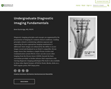
Diagnostic Imaging principles and concepts are augmented by the presentation of images for common clinical conditions. Guiding principles related to minimizing radiation exposure and requesting the most appropriate imaging examination are addressed.Static images are enhanced by the ability to access images stored and displayed on an Html-5 compatible, Dicom image viewer that simulates a simple Picture Archive and Communication system (PACS). Users can also access other imaging from the Dicom viewer (ODIN), beyond the basic curriculum provided, to further advance their experience with viewing diagnostic imaging pathologies.This book is also available in three other digital formats: ePUB (for Nook, iBooks, Kobo etc.), PDF (regular print), PDF (large print).

Word Count: 81603

ISBN: 978-0-88880-611-6

(Note: This resource's metadata has been created automatically by reformatting and/or combining the information that the author initially provided as part of a bulk import process.)
Subject:
Applied Science
Health, Medicine and Nursing
Material Type:
Textbook
Provider:
University of Saskatchewan
Author:
Brent Burbridge
Date Added:
09/30/2017
Undergraduate Diagnostic Imaging Fundamentals
Conditional Remix & Share Permitted
CC BY-NC-SA
Rating
0.0 stars

Short Description:
NewParaDiagnostic Imaging principles and concepts are augmented by the presentation of imaging for common clinical conditions. Guiding principles related to minimizing radiation exposure and requesting the most appropriate imaging examination are addressed. Static images are enhanced by the ability to access images stored and displayed on an html-5 compatible, Dicom image viewer that simulates a simple Picture Archive and Communication system (PACS). Users can also access other imaging from the Dicom viewer (ODIN), beyond the basic curriculum provided, to further advance their experience with viewing diagnostic imaging pathologies.NewParaThis book is also available in three other digital formats: ePUB (for Nook, iBooks, Kobo etc.), PDF (regular print), PDF (large print)

Word Count: 80887

(Note: This resource's metadata has been created automatically by reformatting and/or combining the information that the author initially provided as part of a bulk import process.)

Subject:
Applied Science
Health, Medicine and Nursing
Material Type:
Textbook
Provider:
University of Saskatchewan
Date Added:
09/30/2017
Using Stress and Strain to Detect Cancer!
Read the Fine Print
Educational Use
Rating
0.0 stars

Students are presented with a biomedical engineering challenge: Breast cancer is the second-leading cause of cancer-related death among women and the American Cancer Society says mammography is the best early-detection tool available. Despite this, many women choose not to have them; of all American women at or over age 40, only 54.9% have had a mammogram within the past year. One reason women skip annual mammograms is pain, with 90% reporting discomfort. Is there a way to detect the presence of tumors that is not as painful as mammography but more reliable and quantifiable than breast self-exams or clinical breast exams? This three lesson/three activity unit is designed for first-year accelerated or AP physics classes. It provide hands-on activities to teach the concepts of stress, strain and Hooke's law, which students apply to solve the challenge problem.

Subject:
Applied Science
Engineering
Health, Medicine and Nursing
Material Type:
Full Course
Unit of Study
Provider:
TeachEngineering
Provider Set:
TeachEngineering
Author:
Luke Diamond
Date Added:
09/18/2014
fMRI Bootcamp
Conditional Remix & Share Permitted
CC BY-NC-SA
Rating
0.0 stars

This video lecture series begins with an introduction to the basics of anatomical and functional MRI and the time course of the fMRI signal, then delves into several methods for analyzing fMRI data. The series emphasizes how to think about fMRI data and the steps of analysis rather than the technical execution of each step.
Lecture Topics:

fMRI Bootcamp Part 1 - Basics of fMRI
fMRI Bootcamp Part 2 - fMRI timecourse
fMRI Bootcamp Part 3 - Univariate analysis
fMRI Bootcamp Part 4 - Multivariate analysis
fMRI Bootcamp Part 5 - Multivoxel pattern analysis (MVPA)
fMRI Bootcamp Part 6 - Classification 
fMRI Bootcamp Part 7 - Representational similarity
fMRI Bootcamp Part 8 - fMRI & multiple comparisons
fMRI Bootcamp Part 9 - Hyperalignment

Additional Resources:

Rebecca Saxe’s Lab website
Analyzing fMRI Data page in Modeling and Data Analysis Tools and Datasets
Poldrack R. A., Mumford J. A., Nichols T. E. (2011) Handbook of Functional MRI Data Analysis, Cambridge University Press. ISBN: 9780521517669 [hardcover, eBook, Google Books preview]

Subject:
Applied Science
Health, Medicine and Nursing
Life Science
Physical Science
Material Type:
Full Course
Provider:
MIT
Provider Set:
MIT OpenCourseWare
Author:
Saxe, Rebecca
Date Added:
09/01/2017