This resource is a video abstract of a research paper created by …
This resource is a video abstract of a research paper created by Research Square on behalf of its authors. It provides a synopsis that's easy to understand, and can be used to introduce the topics it covers to students, researchers, and the general public. The video's transcript is also provided in full, with a portion provided below for preview:
"Clinicians use a diversity of anesthetic drugs to regulate memory formation, the perception of pain, and other aspects of consciousness during otherwise painful, unpleasant, or anxiety-provoking experiences. These drugs are well known to vary in their effects on human behavior, but the neural processes in the brain that form the basis of this variation are now being uncovered with functional neuroimaging. In a new study published in the journal _Anesthesiology_, researchers compared the effects of midazolam and ketamine. These two commonly used anesthetics differ in their effects on memory formation, pain perception, and the regions of the brain involved in these processes. While inside an MRI scanner, 26 healthy volunteers received a saline infusion and were asked simple “yes-or-no” questions about a series of spoken words, one-third of which were immediately followed by a painful shock..."
The rest of the transcript, along with a link to the research itself, is available on the resource itself.
This resource is a video abstract of a research paper created by …
This resource is a video abstract of a research paper created by Research Square on behalf of its authors. It provides a synopsis that's easy to understand, and can be used to introduce the topics it covers to students, researchers, and the general public. The video's transcript is also provided in full, with a portion provided below for preview:
"Scientists have uncovered new information on how sensory processing can change in multiple sclerosis, or MS. Their findings could open novel avenues for understanding and treating the disease. People with MS typically experience deficits in their ability to smell and taste, abnormal temperature processing, and heightened sensations of pain or fatigue. But there’s no clear neurocognitive mechanism to explain such diverse symptoms. A new report in the journal Human Brain Mapping suggests these changes may stem from a problem with interoception. Interoception is a lesser-known skill that helps people feel signals originating from inside of the body – such as the heart beating or the digestive system signaling hunger. Many of the sensory processing issues that characterize MS are associated with brain pathways related to interoception. This link prompted researchers to examine how these pathways are affected in the context of the disease..."
The rest of the transcript, along with a link to the research itself, is available on the resource itself.
This course explores the social relevance of neuroscience, considering how emerging areas …
This course explores the social relevance of neuroscience, considering how emerging areas of brain research at once reflect and reshape social attitudes and agendas. Topics include brain imaging and popular media; neuroscience of empathy, trust, and moral reasoning; new fields of neuroeconomics and neuromarketing; ethical implications of neurotechnologies such as cognitive enhancement pharmaceuticals; neuroscience in the courtroom; and neuroscientific recasting of social problems such as addiction and violence. Guest lectures by neuroscientists, class discussion, and weekly readings in neuroscience, popular media, and science studies.
22.56J aims to give graduate students and advanced undergraduates background in the …
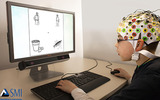
22.56J aims to give graduate students and advanced undergraduates background in the theory and application of noninvasive imaging methods to biology and medicine, with emphasis on neuroimaging. The course focuses on the modalities most frequently used in scientific research (X-ray CT, PET/SPECT, MRI, and optical imaging), and includes discussion of molecular imaging approaches used in conjunction with these scanning methods. Lectures by the professor will be supplemented by in-class discussions of problems in research, and hands-on demonstrations of imaging systems.
This resource is a video abstract of a research paper created by …
This resource is a video abstract of a research paper created by Research Square on behalf of its authors. It provides a synopsis that's easy to understand, and can be used to introduce the topics it covers to students, researchers, and the general public. The video's transcript is also provided in full, with a portion provided below for preview:
"The timing of return to play is one of the most critical decisions made following ACL reconstruction. Returning too early carries the risk of graft failure, while returning too late could cost patients financial and athletic opportunities. Unfortunately, no universal, objective method exists to determine the best time for athletes to resume full activities. But according to a new study reported in The American Journal of Sports Medicine, such methods could be on the horizon, with the help of MRI technology. One promising marker for estimating the best time for athletes to return to play is ACL graft maturation. That’s the process by which a surgical graft makes the transformation from tendon tissue into a substance similar to a normal ACL. The problem is that this transformation is difficult to track over time. Tissue biopsies are currently the gold standard, but are invasive and, in most cases, impractical to perform..."
The rest of the transcript, along with a link to the research itself, is available on the resource itself.
This resource is a video abstract of a research paper created by …
This resource is a video abstract of a research paper created by Research Square on behalf of its authors. It provides a synopsis that's easy to understand, and can be used to introduce the topics it covers to students, researchers, and the general public. The video's transcript is also provided in full, with a portion provided below for preview:
"Throwing injuries among Little League players are currently on the rise. While studies have evaluated the risk factors for throwing injury among these competitive young athletes, no study to date has resorted to serial MRI among asymptomatic Little League players over time. Using this technique, researchers have now discovered several risk factors that predispose youth baseball players to elbow injuries. The team examined the dominant and non-dominant elbows of 26 Little League baseball players. Before the study, each player reported no symptoms of injury and had undergone both pre- and postseason MRI 3 years prior. In addition to conducting repeat elbow MRI at their 3-year follow-up visit, the researchers assessed the players’ shoulders and elbows, throwing history, playing status, and arm pain. MRI results revealed abnormalities in the dominant elbow of 15 players—more than half of the cohort..."
The rest of the transcript, along with a link to the research itself, is available on the resource itself.
Psychology is designed to meet scope and sequence requirements for the single-semester …
Psychology is designed to meet scope and sequence requirements for the single-semester introduction to psychology course. The book offers a comprehensive treatment of core concepts, grounded in both classic studies and current and emerging research. The text also includes coverage of the DSM-5 in examinations of psychological disorders. Psychology incorporates discussions that reflect the diversity within the discipline, as well as the diversity of cultures and communities across the globe.Senior Contributing AuthorsRose M. Spielman, Formerly of Quinnipiac UniversityContributing AuthorsKathryn Dumper, Bainbridge State CollegeWilliam Jenkins, Mercer UniversityArlene Lacombe, Saint Joseph's UniversityMarilyn Lovett, Livingstone CollegeMarion Perlmutter, University of Michigan
By the end of this section, you will be able to:Explain the …
By the end of this section, you will be able to:Explain the functions of the spinal cordIdentify the hemispheres and lobes of the brainDescribe the types of techniques available to clinicians and researchers to image or scan the brain
This resource is a video abstract of a research paper created by …
This resource is a video abstract of a research paper created by Research Square on behalf of its authors. It provides a synopsis that's easy to understand, and can be used to introduce the topics it covers to students, researchers, and the general public. The video's transcript is also provided in full, with a portion provided below for preview:
"ACL injuries are common among athletes of all types. Often, they’re associated with ramp lesions—injuries to the medial meniscus, which cushions the femur and provides stability to the knee joint. Unfortunately, ramp lesions often go undiagnosed, and the most appropriate course of treatment is the subject of much debate. To bridge this gap, researchers examined the prevalence and characteristics of ramp lesions in patients undergoing ACL reconstruction. Their findings are reported in _The American Journal of Sports Medicine_. The team retrospectively evaluated all patients who underwent ACL reconstruction at a hospital in Lyon, France, between November 2015 and November 2018. They used a combination of imaging and surgical techniques to identify whether ramp lesions were present..."
The rest of the transcript, along with a link to the research itself, is available on the resource itself.
This resource is a video abstract of a research paper created by …
This resource is a video abstract of a research paper created by Research Square on behalf of its authors. It provides a synopsis that's easy to understand, and can be used to introduce the topics it covers to students, researchers, and the general public. The video's transcript is also provided in full, with a portion provided below for preview:
"New research reported in the American Journal of Sports Medicine suggests that a resorbable polyurethane implant could help patients recover from meniscal deficiency. Researchers showed that the implant was able to improve knee function and reduce pain in patients with partial meniscal defects. Meniscal lesions are the most common injury requiring orthopedic surgery today. In Europe, surgeons perform 400,000 such procedures each year—and in the US, more than a million. Removing the damaged tissue used to be the preferred method for treating meniscal tears. But given the risk of developing arthritis in the absence of this vital cushion, surgeons are now opting to repair or even regenerate the injured tissue. To determine how viable regeneration might be, researchers examined 155 patients fitted with a polyurethane scaffold designed to promote tissue regrowth. One hundred forty-one of these patients previously had part of their meniscus removed and suffered from post-surgical pain..."
The rest of the transcript, along with a link to the research itself, is available on the resource itself.
Is it a tumor? Magnetic Resonance Imaging (MRI) can tell. Your head …
Is it a tumor? Magnetic Resonance Imaging (MRI) can tell. Your head is full of tiny radio transmitters (the nuclear spins of the hydrogen nuclei of your water molecules). In an MRI unit, these little radios can be made to broadcast their positions, giving a detailed picture of the inside of your head.
This resource is a video abstract of a research paper created by …
This resource is a video abstract of a research paper created by Research Square on behalf of its authors. It provides a synopsis that's easy to understand, and can be used to introduce the topics it covers to students, researchers, and the general public. The video's transcript is also provided in full, with a portion provided below for preview:
"Knee problems such as instability of the kneecap and arthritis can be telltale signs that it’s time for surgery. But they’re not the only ones. A new study suggests that focal lesions of the cartilage that cushions the kneecap, or patella, and of the trochlea, the groove where the patella rests, are just as important and can have similar causes. Understanding how these lesions are related to knee alignment and morphology could help clinicians develop more tailored and durable treatments. Researchers reached that conclusion after comparing the knee anatomy of 135 patients with focal patellofemoral cartilage lesions, but no arthritis, to that of 100 patients with normal cartilage. Using M-R-I, they assessed features including patellar morphology, trochlear morphology, patellar malalignment, and the quadriceps vector. Patients with patellofemoral lesions showed greater trochlear dysplasia and patellar malalignment than those in the control group..."
The rest of the transcript, along with a link to the research itself, is available on the resource itself.
This resource is a video abstract of a research paper created by …
This resource is a video abstract of a research paper created by Research Square on behalf of its authors. It provides a synopsis that's easy to understand, and can be used to introduce the topics it covers to students, researchers, and the general public. The video's transcript is also provided in full, with a portion provided below for preview:
"Rotator cuff injuries are the most common form of injury to the shoulder. In the case of large and massive, irreparable rotator cuff tears, the gold standard treatment is a partial or maximal repair. Despite short-term pain relief and improved function, however, re-tear rates following maximal repair can range from 50 to 90 percent. That makes it important to explore alternative treatment approaches. In a new study reported in the_ American Journal of Sports of Medicine_, researchers compared maximal repair to maximal with bridging interpositional dermal allograft in patients with massive rotator cuff tears. Maximal repair recreates the rotator cable much like a suspension bridge to regain rotator cuff function. Bridging interpositional allograft, on the other hand, uses tissue grafts to bridge bone to tendon. Thirty patients with massive, retracted, and irreparable rotator cuff tears were randomly allocated to receive either procedure..."
The rest of the transcript, along with a link to the research itself, is available on the resource itself.
This resource is a video abstract of a research paper created by …
This resource is a video abstract of a research paper created by Research Square on behalf of its authors. It provides a synopsis that's easy to understand, and can be used to introduce the topics it covers to students, researchers, and the general public. The video's transcript is also provided in full, with a portion provided below for preview:
"Shoulder dislocations and partial dislocations are some of the most common and disabling injuries in active young people, especially athletes. After the bone has been popped back and the initial agony is over, the issue of what to do next is much debated. One option is surgery to try to repair the damage, but whether that is preferable to less-invasive care remains controversial. Researchers now have evidence that supports the surgical option, at least for young people suffering a first event. The team monitored the degree of bone damage to the shoulder socket, or glenoid bone, which holds the head of the humerus in place. Until now, there has not been much information about the extent of this damage to guide a decision on the best approach. The shoulders of 714 student athletes were imaged by MRI scanning, and the subjects were then followed for four years..."
The rest of the transcript, along with a link to the research itself, is available on the resource itself.
Systems Neuroscience Laboratory consists of a series of laboratories designed to give …
Systems Neuroscience Laboratory consists of a series of laboratories designed to give students experience with basic techniques for conducting systems neuroscience research. It includes sessions on anatomical, neurophysiological, and data acquisition and analysis techniques, and the ways these techniques are used to study nervous system function. Training is provided in the art of scientific writing with feedback designed to improve writing skills. Assignments include weekly preparation for lab sessions, two major research reports and a series of basic computer programming tutorials (MATLAB). The class involves the use of experimental animals. Enrollment is limited.
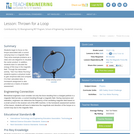
In this lesson, students begin to focus on the torque associated with …
In this lesson, students begin to focus on the torque associated with a current carrying loop in a magnetic field. Students are prompted with example problems and use diagrams to visualize the vector product. In addition, students learn to calculate the energy of this loop in the magnetic field. Several example problems are included and completed as a class. A homework assignment is also attached as a means of student assessment.
This resource is a video abstract of a research paper created by …
This resource is a video abstract of a research paper created by Research Square on behalf of its authors. It provides a synopsis that's easy to understand, and can be used to introduce the topics it covers to students, researchers, and the general public. The video's transcript is also provided in full, with a portion provided below for preview:
"A new report in The American Journal of Sports Medicine sheds light on the anatomic features that predispose certain individuals to develop hip impingement, helping answer a question that’s perplexed orthopedic specialists for years. Femoroacetabular impingement, or FAI, occurs when extra bone grows along the hip, causing friction in the joint that can lead to symptoms ranging from pain to premature osteoarthritis. What’s perplexing is that many individuals with bone deformities assumed to be linked to FAI never go on to develop symptoms of the disorder. That’s made it difficult to distinguish what aspects of joint morphology actually give rise to FAI. To uncover the factors that may predict a symptomatic state, an international team of researchers turned to three-dimensional magnetic resonance imaging, which they used to examine the interplay between hip anatomy and various factors relating to the pelvis and also to the spine, that are termed spinopelvic parameters..."
The rest of the transcript, along with a link to the research itself, is available on the resource itself.
In this activity, students take the age old concept of etch-a-sketch a …
In this activity, students take the age old concept of etch-a-sketch a step further. Using iron filings, students begin visualizing magnetic field lines. To do so, students use a compass to read the direction of the magnet's magnetic field. Then, students observe the behavior of iron filings near that magnet as they rotate the filings about the magnet. Finally, students study the behavior of iron filings suspended in mineral oil which displays the magnetic field in three dimensions.
No restrictions on your remixing, redistributing, or making derivative works. Give credit to the author, as required.
Your remixing, redistributing, or making derivatives works comes with some restrictions, including how it is shared.
Your redistributing comes with some restrictions. Do not remix or make derivative works.
Most restrictive license type. Prohibits most uses, sharing, and any changes.
Copyrighted materials, available under Fair Use and the TEACH Act for US-based educators, or other custom arrangements. Go to the resource provider to see their individual restrictions.