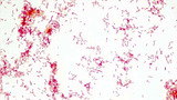
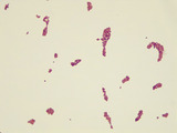
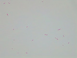
This micrograph was taken at 1000X total magnifcation on a brightfield microscope. …
This micrograph was taken at 1000X total magnifcation on a brightfield microscope. The subject is Escherichia coli cells grown in broth culture overnight at 37 degrees Celsius. The cells were heat-fixed to a slide and stained for 1 minute with safranin red stain prior to visualization.Image credit: Emily Fox
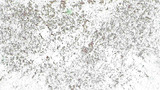
This micrograph was taken at 100X total magnifcation on a brightfield microscope. …
This micrograph was taken at 100X total magnifcation on a brightfield microscope. The subject is Escherichia coli cells grown in broth culture overnight at 37 degrees Celsius. The cells were heat-fixed to a slide and stained for 1 minute with safranin red stain prior to visualization.Image credit: Emily Fox
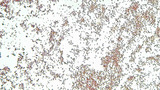
This micrograph was taken at 400X total magnifcation on a brightfield microscope. …
This micrograph was taken at 400X total magnifcation on a brightfield microscope. The subject is Escherichia coli cells grown in broth culture overnight at 37 degrees Celsius. The cells were heat-fixed to a slide and stained for 1 minute with safranin red stain prior to visualization.Image credit: Emily Fox
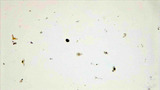
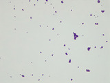
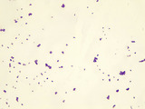
This micrograph was taken at 1000X total magnifcation on a brightfield microscope. …
This micrograph was taken at 1000X total magnifcation on a brightfield microscope. The subject is Lactococcus lactis cells grown on agar at 37 degrees Celsius. The cells were heat-fixed to a slide and Gram stained prior to visualization.Image credit: Emily Fox
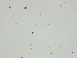
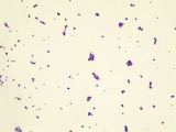
This micrograph was taken at 1000X total magnifcation on a brightfield microscope. …
This micrograph was taken at 1000X total magnifcation on a brightfield microscope. The subject is Micrococcus cells grown on nutrient agar at 25 degrees Celsius. The cells were heat-fixed to a slide and Gram stained prior to visualization.Image credit: Emily Fox
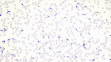
This micrograph was taken at 1000X total magnifcation on a brightfield microscope. …
This micrograph was taken at 1000X total magnifcation on a brightfield microscope. The subject is Micrococcus luteus cells grown on agar at 37 degrees Celsius. The cells were heat-fixed to a slide and Gram stained prior to visualization.Image credit: Emily Fox
This micrograph was taken at 1000X total magnifcation on a brightfield microscope. …
This micrograph was taken at 1000X total magnifcation on a brightfield microscope. The subject is Micrococcus luteus cells grown on nutrient agar plates at 37 degrees Celsius. The cells were stained in a smear of nigrosin negative stain prior to visualization.Image credit: Emily Fox
This micrograph was taken at 1000X total magnifcation on a brightfield microscope. …
This micrograph was taken at 1000X total magnifcation on a brightfield microscope. The subject is Neisseria sicca cells grown on nutrient agar at 37 degrees Celsius. The cells were heat-fixed to a slide and Gram stained prior to visualization.Image credit: Emily Fox
This micrograph was taken at 1000X total magnifcation on a brightfield microscope. …
This micrograph was taken at 1000X total magnifcation on a brightfield microscope. The subject is Pseudomonas putida cells grown in broth culture overnight at 37 degrees Celsius. The cells were heat-fixed to a slide and Gram stained prior to visualization.Image credit: Emily Fox
This micrograph was taken at 1000X total magnifcation on a brightfield microscope. …
This micrograph was taken at 1000X total magnifcation on a brightfield microscope. The subject is Staphylococcus aureus cells grown on nutrient agar at 37 degrees Celsius. The cells were heat-fixed to a slide and Gram stained prior to visualization.Image credit: Emily Fox
This micrograph was taken at 400X total magnifcation on a brightfield microscope. …
This micrograph was taken at 400X total magnifcation on a brightfield microscope. The subject is giemsa-stained Trypanosoma cruzi in a blood smear.Image credit: Emily Fox
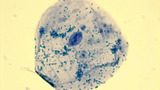
This micrograph was taken at 1000X total magnifcation on a brightfield microscope. …
This micrograph was taken at 1000X total magnifcation on a brightfield microscope. The subject is human cheek epithelial cells collected fresh with a toothpick. The cells were stained with methylene blue stain prior to visualization.Image credit: Emily Fox
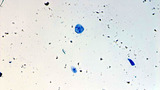
This micrograph was taken at 100X total magnifcation on a brightfield microscope. …
This micrograph was taken at 100X total magnifcation on a brightfield microscope. The subject is human cheek epithelial cells collected fresh with a toothpick. The cells were stained with methylene blue stain prior to visualization.Image credit: Emily Fox
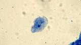
This micrograph was taken at 400X total magnifcation on a brightfield microscope. …
This micrograph was taken at 400X total magnifcation on a brightfield microscope. The subject is human cheek epithelial cells collected fresh with a toothpick. The cells were stained with methylene blue stain prior to visualization.Image credit: Emily Fox
After watching a short online video that recaps the enormous scale of …
After watching a short online video that recaps the enormous scale of accumulating plastic waste in our oceans, student teams are challenged to devise a method to remove the most plastic microbeads from a provided commercial personal care product—such as a facial cleanser or body wash. They brainstorm filtering methods ideas and design their own specific procedures that use teacher-provided supplies (coffee filters, funnels, plastic syringes, vinyl tubing, water, plastic bags) to extract the microplastics as efficiently as possible. The research and development student teams compare the final masses of their extracted microbeads to see which filter solutions worked best. Students suggest possible future improvements to their filter designs. A student worksheet is provided.
This resource is a video abstract of a research paper created by …
This resource is a video abstract of a research paper created by Research Square on behalf of its authors. It provides a synopsis that's easy to understand, and can be used to introduce the topics it covers to students, researchers, and the general public. The video's transcript is also provided in full, with a portion provided below for preview:
"Reproducibility is extremely important in science. But no matter how much effort is put into standardizing protocols, small differences seem inevitable in the way experiments are performed among laboratories. These small differences can add up to discrepancies that complicate data interpretation. One field where this issue looms large is neuroscience – particularly in experiments involving histological sections of the mouse brain. Such studies require correct identification of specific brain regions for accurate interpretation of results. But the mouse brain is small and complex. Brain atlases can be invaluable in mapping, but applying this information in laboratory experiments is difficult. A new automated system aims to solve this problem by taking the guesswork – and potential observer error and bias – out of the equation. Much like a GPS system in a car, the program – called NeuroInfo – helps researchers navigate through the microscopic anatomy of a brain section..."
The rest of the transcript, along with a link to the research itself, is available on the resource itself.
Students are introduced to the growing worldwide environmental problems that stem from …
Students are introduced to the growing worldwide environmental problems that stem from plastic waste. What they learn about microplastics and the typical components of the U.S. water treatment process prepares them to conduct three engaging associated activities. During the lesson, students become more aware of the pervasiveness and value of plastic as well as the downstream pollution and health dangers. They learn how plastic materials don’t go away, but become microplastic pollution that accumulates in water resources as well as human and other animal bodies. They examine their own plastic use, focusing on what they discard daily, and think about better ways to produce or package those items to eliminate or reduce their likelihood of ending up as microplastic pollution. A concluding writing assignment reveals their depth of comprehension. The lesson is enhanced by arranging for a local water treatment plant representative to visit the class for Qs and As. In three associated activities, students design/test microplastic particle filtering methods for commercial products, create mini wastewater treatment plant working models that remove waste and reclaim resources from simulated wastewater, and design experiments to identify the impact of microplastics on micro-invertebrates.
No restrictions on your remixing, redistributing, or making derivative works. Give credit to the author, as required.
Your remixing, redistributing, or making derivatives works comes with some restrictions, including how it is shared.
Your redistributing comes with some restrictions. Do not remix or make derivative works.
Most restrictive license type. Prohibits most uses, sharing, and any changes.
Copyrighted materials, available under Fair Use and the TEACH Act for US-based educators, or other custom arrangements. Go to the resource provider to see their individual restrictions.