(This case study was added to OER Commons as one of a …
(This case study was added to OER Commons as one of a batch of over 700. It has relevant information which may include medical imagery, lab results, and history where relevant. A link to the final diagnosis can be found at the end of the case study for review. The first paragraph of the case study -- typically, but not always the clinical presentation -- is provided below.)
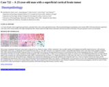
A 23-year-old male with no significant past history, presented with a tonic-clonic generalized seizure. Physical and neurological examinations were normal. MRI of the brain showed a superficial cystic mass with a mural nodule in the right frontoparietal lobe. Patient underwent right frontal craniotomy with total resection of the tumor. The post-operative course was uneventful.
(This case study was added to OER Commons as one of a …
(This case study was added to OER Commons as one of a batch of over 700. It has relevant information which may include medical imagery, lab results, and history where relevant. A link to the final diagnosis can be found at the end of the case study for review. The first paragraph of the case study -- typically, but not always the clinical presentation -- is provided below.)
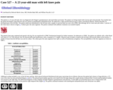
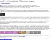
The patient is a 23 year-old male who was diagnosed with Wegner's granulomatosis and renal failure previously. The patient was being treated with Cytoxan and corticosteroids. Four months later, the patient developed a lung infection and was started on trimethoprim-sulfamethoxazole. He underwent a video-assisted thoracoscopy to drain a left pleural effusion. Shortly thereafter, he developed pain in his left knee. The patient underwent debridement of the left knee and a sample of synovial fluid was sent to the UPMC Microbiology lab for Gram stain and culture. A picture of the Gram stain is shown in figure 1. Cultures grew the same organism. The patient continued with antibiotic treatment at an outside hospital.
(This case study was added to OER Commons as one of a …
(This case study was added to OER Commons as one of a batch of over 700. It has relevant information which may include medical imagery, lab results, and history where relevant. A link to the final diagnosis can be found at the end of the case study for review. The first paragraph of the case study -- typically, but not always the clinical presentation -- is provided below.)
A 24-year-old Caucasian man was admitted following a 10-day history of severe headache leading to collapse on the street. On presentation, he was confused and agitated with left-sided weakness and a positive left Babinski sign. His medical history was significant for asthma and non-Hodgkin's lymphoma 10 years previously treated with chemotherapy and radiation therapy with no recurrence.
(This case study was added to OER Commons as one of a …
(This case study was added to OER Commons as one of a batch of over 700. It has relevant information which may include medical imagery, lab results, and history where relevant. A link to the final diagnosis can be found at the end of the case study for review. The first paragraph of the case study -- typically, but not always the clinical presentation -- is provided below.)
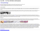
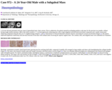
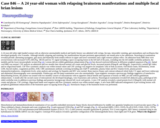
A 24-year-old man presented with new onset of generalized tonic clonic seizure. Prior to admission, the patient reported an enlarging painless scalp mass for the previous two months, without having sought medical attention. MRI of the brain revealed a 7.7 cm heterogeneously enhancing mass with a large subgaleal extracalvarial/extracranial component and a smaller intracranial enhancing component along the dura on T1 weighted images (Figures 1 and 2). Noncontrast CT showed abnormal calvarial sclerosis concerning for osseous involvement (Figure 3). The patient underwent biparietal craniectomy and sub-total resection of the mass with residual tumor involving the superior sagittal sinus. The cranium, dura, and subgaleal mass were submitted for evaluation.
(This case study was added to OER Commons as one of a …
(This case study was added to OER Commons as one of a batch of over 700. It has relevant information which may include medical imagery, lab results, and history where relevant. A link to the final diagnosis can be found at the end of the case study for review. The first paragraph of the case study -- typically, but not always the clinical presentation -- is provided below.)
The mother of this fetus was a 24 year old G1 P0 woman with multiple medical problems. These include cerebral palsy, mixed hearing loss, a history of seizures, history of a heart murmur, surgery for cleft lip, and asthma. The current pregnancy was without complications through the first two trimesters. An 18 week anatomy scan was unremarkable. A fetal echocardiogram at 18 weeks was also unremarkable. Early in the third trimester, a clinical concern for size less than dates arose. An ultrasound at 30 weeks found an enlarged fetal heart. No evidence of hydrops was identified, however. On a routine clinic visit at 32 2/7 weeks gestation, the mother complained of no fetal movement for the previous two days. A bedside ultrasound confirmed an intrauterine fetal demise. Labor was induced, with delivery of the stillborn female fetus.
(This case study was added to OER Commons as one of a …
(This case study was added to OER Commons as one of a batch of over 700. It has relevant information which may include medical imagery, lab results, and history where relevant. A link to the final diagnosis can be found at the end of the case study for review. The first paragraph of the case study -- typically, but not always the clinical presentation -- is provided below.)
A previously healthy 24-year-old white female patient presented to the emergency room with back pain. The pain, which had been ongoing for the past four days, started in her lower back and wrapped around her left leg down below the knee. It was constant and worsened with any activity. Bladder and bowel function were unaffected. The patient denied any prior history of back pain or back injury. She did not have any history of trauma. On physical examination, no focal neurological findings were noted.
(This case study was added to OER Commons as one of a …
(This case study was added to OER Commons as one of a batch of over 700. It has relevant information which may include medical imagery, lab results, and history where relevant. A link to the final diagnosis can be found at the end of the case study for review. The first paragraph of the case study -- typically, but not always the clinical presentation -- is provided below.)
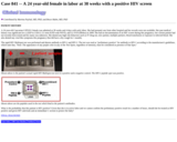
The rapid HIV Multispot test was performed and detests antibody to HIV-1 and HIV-2. The test was read as "preliminary positive" for antibody to HIV-1 according to the manufacturer's guidelines, which state that, "Note: The appearance of any purple color in any of the Test Spots, regardless of intensity, must be considered as presence of that Spot."
(This case study was added to OER Commons as one of a …
(This case study was added to OER Commons as one of a batch of over 700. It has relevant information which may include medical imagery, lab results, and history where relevant. A link to the final diagnosis can be found at the end of the case study for review. The first paragraph of the case study -- typically, but not always the clinical presentation -- is provided below.)
The patient reports past medical history of nephrolithiasis only. The patient denies prior surgical procedures. She denies family history of colon, uterine, breast and ovarian carcinomas. Relevant obstetric-gynecological history consists of G3P2012, two full term spontaneous vaginal deliveries. The patient has no history of sexually transmitted infectious diseases and is currently sexually active, taking oral contraceptive pills. No history of abnormal cervical PAP smears.
(This case study was added to OER Commons as one of a …
(This case study was added to OER Commons as one of a batch of over 700. It has relevant information which may include medical imagery, lab results, and history where relevant. A link to the final diagnosis can be found at the end of the case study for review. The first paragraph of the case study -- typically, but not always the clinical presentation -- is provided below.)
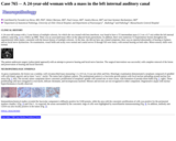
A 24-year-old woman with a 3 year history of multiple sclerosis, for which she was treated with beta interferon, was found to have a T2 intermediate mass (1.3 cm x 0.7 cm) within the left internal auditory canal (Fig. 1) in a follow up MRI. There was no associated mass effect on the adjacent brain parenchyma. In addition, there were numerous T2 hyperintense lesions throughout the supratentorial white matter, consistent with the known history of multiple sclerosis. At the time, she did not have any related symptoms; there was no reported abnormality of hearing or balance, and no facial nerve dysfunction. On examination, visual fields and acuity were normal and cranial nerves II through XII were intact, with normal hearing on both sides. Motor-sensory skills were normal.
(This case study was added to OER Commons as one of a …
(This case study was added to OER Commons as one of a batch of over 700. It has relevant information which may include medical imagery, lab results, and history where relevant. A link to the final diagnosis can be found at the end of the case study for review. The first paragraph of the case study -- typically, but not always the clinical presentation -- is provided below.)
A 24 year-old white right handed woman with an otherwise unremarkable medical and family history was admitted with vertigo, hiccups, intractable vomiting, gait unsteadiness and oscillopsia that developed gradually over 9 months. Although initially relapsing and remitting, her manifestations became persistent approximately one month prior to her admission. Neurological examination revealed periodic alternating nystagmus, gait ataxia and bilaterally brisk tendon reflexes in upper and lower extremities and a right extensor plantar reflex. On admission brain MRI there were several lesions with increased T2/FLAIR (Fig. 1BCD) and low T1 signal including a space-occupying lesion in the left half of the pons, extending into the left middle cerebellar peduncle, the medulla and the lower mesencephalic tectum (Fig.1A). Lesions did not exhibit gadolinium enhancement (Fig.1A) but showed restricted diffusion in diffusion-weighted sequences (Fig.1E). Spinal cord MRI was normal. CSF analysis on admission and two months later showed normal cell counts and glucose levels, increased protein (83mg/dL and 90mg/dL, respectively), normal IgG index and no oligoclonal bands. CSF flow cytometric analysis was within normal values and CSF cytology was negative for neoplastic cells in both occasions. Full blood counts, biochemistry, serum LDH, thyroid studies, coagulation profiles, ESR, CRP, autoimmune antibody screening including anti-AQP IV antibodies, plasma folate and B12 vitamin levels, plasma protein immunoelectrophoresis, complement levels, urine analysis and serology for hepatitis and HIV were normal. Bone marrow biopsy and contrast-enhanced whole body CT, upper and lower endoscopy and abdominal ultrasonography were unremarkable. Fundoscopy and slit-lamp examination were also unremarkable. Upon magnetic resonance spectroscopy findings suggestive of tumefactive demyelinating lesions, the patient was treated with two monthly courses of mitoxantrone with no apparent clinical benefit and expansion of lesions on MRI, yet still without gadolinium enhancement. A month after the second course of mitoxantrone she developed dysphagia, voice hoarseness and cachexia. Neurological examination revealed left abducens palsy, pathological left-sided cerebellar tests in addition to previous findings and posterior laryngoscopy showed left vocal cord paresis. A new CSF analysis revealed a raised protein level (130mg/dL) with normal cell counts and glucose. CSF cytology, oligoclonal bands were negative and flow cytometric analysis was within normal ranges. A new brain MRI indicated enlargement of pre-existing lesions with peripheral gadolinium enhancement of the brainstem space-occupying lesion (Fig.1F). Stereotactic biopsy of the brainstem lesion was performed.
(This case study was added to OER Commons as one of a …
(This case study was added to OER Commons as one of a batch of over 700. It has relevant information which may include medical imagery, lab results, and history where relevant. A link to the final diagnosis can be found at the end of the case study for review. The first paragraph of the case study -- typically, but not always the clinical presentation -- is provided below.)
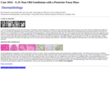
A 25-year-old gentleman presented with short history of headache and gait disturbances. Radiology revealed a heterogeneously enhancing mass in the fourth ventricle extending from midbrain to pons. CT scan showed a hyperdense mass in the 4th ventricle. There was no calcification or cysts seen within the lesion. MRI showed a well defined mass in the fourth ventricle. The lesion was isointense on T1, hyperintense on T2 (Fig. 1A) and enhanced heterogeneously on contrast administration (Fig. 1B). The contrast images showed tumor infiltration into brain stem suggesting a poor plane of cleavage, better appreciated on sagittal MRI (Fig. 1C). FLAIR MRI showed slightly hyperintense lesion suggesting a cellular tumor (Fig. 1D). Though the lesion showed no calcifications or cysts or areas of bleed, the enhancement pattern and infiltration in the fourth ventricular floor suggested a radiological diagnosis of ependymoma. The lesion was operated through a midline suboccipital craniotomy. The lesion was yellow grey, not very vascular and was attached to the brain stem. A thin sliver of tumor adherent to the brain stem was left. Near total resection could be achieved.
(This case study was added to OER Commons as one of a …
(This case study was added to OER Commons as one of a batch of over 700. It has relevant information which may include medical imagery, lab results, and history where relevant. A link to the final diagnosis can be found at the end of the case study for review. The first paragraph of the case study -- typically, but not always the clinical presentation -- is provided below.)
The patient is a 2.5 month old female who presented with weight loss, stridor, hypertonia, and abnormal visual tracking. She was the result of a full term pregnancy with no complications and unremarkable delivery. Of note, the neurological exam at that time was normal. She regained birth weight quickly but had weight gaining difficulties starting in the second month. There were additional developmental delays such as no social smile, no object tracking, nor engaging with environment. There was back arching noted while feeding.
(This case study was added to OER Commons as one of a …
(This case study was added to OER Commons as one of a batch of over 700. It has relevant information which may include medical imagery, lab results, and history where relevant. A link to the final diagnosis can be found at the end of the case study for review. The first paragraph of the case study -- typically, but not always the clinical presentation -- is provided below.)
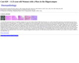
A 25-year-old woman had a history of 4 episodes of epilepsy over two years without treatment. MRI showed that there was a solid circumscribed, hyperintense and nonenhancing tumor (Fig 1) measuring 6x4x4cm in the hippocampus. T1-weighted and T2-weighted scans showed mixed signals without calcification in the tumor and no peritumoral edema. The tumor was totally resected. Macroscopically, the surgical sample was bits and pieces. Histologically, the tumor included glial, neuronal and mixed glioneuronal populations. The spindle-shaped tumor cells showed diffuse growth and displayed characteristic angiocentric arrangements around small parenchymal vessels forming perivascular pseudorosettes. There were single or multilayered tumor cells arranged with ependymal features (Fig 2). The tumor cells of the hippocampal surface formed distinctive rosettes (Fig 3) which have not been previously described. The spindle tumor cells were bipolar with elongated nuclei and inconspicuous nucleoli. In some areas, the tumor cells were schwannoma-like (Fig 4). We could find single neurons interspersed within the tumor tissue. Necrosis and mitotic activity were absence. Immunohistochemistry was positive for GFAP (Fig 5), Vimentin and S-100 and negative for neurofilament protein, Syn, CgA and NeuN. EMA showed "dot-like" positive staining (Fig 6). The proliferation index was less than 1%. Epilepsy disappeared after the operation. We only follow-up four months till now and There was no evidence of recurrence on MRI at a four month follow-up.
(This case study was added to OER Commons as one of a …
(This case study was added to OER Commons as one of a batch of over 700. It has relevant information which may include medical imagery, lab results, and history where relevant. A link to the final diagnosis can be found at the end of the case study for review. The first paragraph of the case study -- typically, but not always the clinical presentation -- is provided below.)
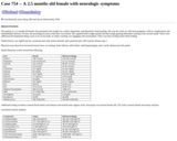
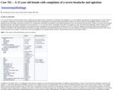
A 25 year old African American female with no significant past medical history presented to the hospital with complaints of a severe headache and agitation for approximately one week. While in the emergency department, she became extremely agitated and violent, requiring physical restraints and benzodiazepine sedation. Her initial workup was unremarkable with the exception of a qualitative urine toxicology screen that was positive for cannabinoids. The patient was admitted to the inpatient psychiatry service for presumed acute psychosis. Within a few hours, she developed generalized seizures and required intubation for airway protection. A CT scan of her head, a brain MRI, and an EEG were negative for any acute intracranial pathology. Despite sedation and anti-epileptic medication, the patient continued to have persistent seizures and agitation. She was immediately transferred to the ICU where her sedation was increased and she was maintained on mechanical ventilation. She remained afebrile, had stable vital signs, and had an unremarkable physical exam. However, any time sedation was held she developed generalized tonic clonic seizure activity and had to be re-sedated. A work-up for acute status epilepticus was initiated, and the results of relevant laboratory tests are provided in Table 1.
(This case study was added to OER Commons as one of a …
(This case study was added to OER Commons as one of a batch of over 700. It has relevant information which may include medical imagery, lab results, and history where relevant. A link to the final diagnosis can be found at the end of the case study for review. The first paragraph of the case study -- typically, but not always the clinical presentation -- is provided below.)
The patient is a 25 year-old male with no significant past medical history. He presented to his primary care physician complaining of a constant, throbbing left parietal headache for four to five days. He also had intermittent, generalized sweating accompanied by chills, fatigue, malaise, myalgias, and arthralgias for two to three days. Ten days prior, the patient had returned from a trip to India. He was born and raised there and visits yearly. He received no vaccinations prior to his trip and was never vaccinated for previous visits. The patient was afebrile with a completely unremarkable physical examination.
(This case study was added to OER Commons as one of a …
(This case study was added to OER Commons as one of a batch of over 700. It has relevant information which may include medical imagery, lab results, and history where relevant. A link to the final diagnosis can be found at the end of the case study for review. The first paragraph of the case study -- typically, but not always the clinical presentation -- is provided below.)
A 25 year-old male presents with high fever (105 degrees F) for approximately one week, accompanied by nausea, vomiting, arthralgias, and myalgias. He reports that he spent the past 15 months serving with the military in Afghanistan and only recently returned. He had been taking mefloquine for malarial prophylaxis but discontinued it several weeks-months ago.
(This case study was added to OER Commons as one of a …
(This case study was added to OER Commons as one of a batch of over 700. It has relevant information which may include medical imagery, lab results, and history where relevant. A link to the final diagnosis can be found at the end of the case study for review. The first paragraph of the case study -- typically, but not always the clinical presentation -- is provided below.)
A 25-year-old white male with a history of schizoaffective disorder was brought to the emergency room in February with a 24-hour history of difficulty breathing and not acting normally.
(This case study was added to OER Commons as one of a …
(This case study was added to OER Commons as one of a batch of over 700. It has relevant information which may include medical imagery, lab results, and history where relevant. A link to the final diagnosis can be found at the end of the case study for review. The first paragraph of the case study -- typically, but not always the clinical presentation -- is provided below.)
A 25-year-old, previously healthy woman presented with a 3-year history of lumbago with radiation to the right lower extremity and was admitted to our hospital. Pain was of mechanical type and responded well to rest. Complete neurological examination was normal. Magnetic resonance imaging (MRI) showed two well-circumscribed isolated intradural masses at levels L2-L3 and S1-S2. One at level L2-L3 measured 3.2 cm in its longest axis. The other extending from S1-S2 measured 2.2 cm in its long axis. Both masses were heterogeneous in T2 and strongly enhanced after gadolinium injection (Figures 1a, 1b,1c). No other lesions were identified elsewhere on craniospinal axis by MRI. Both masses were completely resected. The patient was discharged ten days later. The MRI performed 10 months after surgery showed no recurrence of the tumors.
(This case study was added to OER Commons as one of a …
(This case study was added to OER Commons as one of a batch of over 700. It has relevant information which may include medical imagery, lab results, and history where relevant. A link to the final diagnosis can be found at the end of the case study for review. The first paragraph of the case study -- typically, but not always the clinical presentation -- is provided below.)
A 26 year-old female initially noted blurry vision when looking towards the right. Three months later, she noted diplopia when looking towards the right side. Right abducens nerve palsy was documented by her ophthalmologist. Initial brain MRI performed four months after the onset of blurry vision showed a 4mm (AP) X3mm(transverse)X5mm (craniocaudal) area of enhancement anterior to the pons on the right side (Figure 1). Neurology coordinated an extensive serum screening for inflammatory, infectious, and metabolic causes, all of which came back negative. Two to three months later, she started experiencing daily pressure headaches felt mostly in the occipital and suboccipital regions. The follow-up brain MRI showed the presence of a prepontine/premedullary mass, measuring 23mm (AP) X34mm (transverse) X29mm (craniocaudal) (Figure 2). This mass was mostly solid but presented some cystic necrotic areas. The lesion, believed to be extra-axial, exerted mass effect on the pons and medulla without causing abnormal signal within the brainstem. The mass also caused asymmetry of the fourth ventricle without evidence of obstructive hydrocephalus. At the time of consultation with neurosurgery, approximately 7 months after the onset of her blurry vision, the patient reported difficulty swallowing solids for the last two weeks. Other than abducens nerve palsy, the rest of her physical exam was unremarkable. After review of the images with neuroradiology, the suspected diagnosis was a malignant nerve sheet tumor, most probably arising from the abducens cranial nerve. The investigation was completed with a spine MRI which demonstrated the presence of three subcentimetric enhancing nodules, interpreted as possible schwannomas or meningiomas (Figure 3). A catheter angiogram did not reveal any direct arterial feeder that could be embolized.
(This case study was added to OER Commons as one of a …
(This case study was added to OER Commons as one of a batch of over 700. It has relevant information which may include medical imagery, lab results, and history where relevant. A link to the final diagnosis can be found at the end of the case study for review. The first paragraph of the case study -- typically, but not always the clinical presentation -- is provided below.)
A 27 year old female patient, presented with left hemiparesis. Brain MRI showed multiple cystic ring-enhancing lesions involving the cerebrum and cerebellum (Figure-1A). She has received anti-tuberculous treatment for a recent diagnosis with pulmonary tuberculosis, however, the brain lesions showed with no response. Investigations revealed no other medical conditions. She underwent craniotomy and one of the lesions was excised from the left parietal lobe. It consisted of an oval white to gray soft tissue mass with solid white cut surface measuring 1.2x0.8x0.9cm (Figure-1B).
No restrictions on your remixing, redistributing, or making derivative works. Give credit to the author, as required.
Your remixing, redistributing, or making derivatives works comes with some restrictions, including how it is shared.
Your redistributing comes with some restrictions. Do not remix or make derivative works.
Most restrictive license type. Prohibits most uses, sharing, and any changes.
Copyrighted materials, available under Fair Use and the TEACH Act for US-based educators, or other custom arrangements. Go to the resource provider to see their individual restrictions.