Updating search results...

Search Resources

220 Results

View
Selected filters:
  • Veterinary Medicine
Abomasum - Anatomy & Physiology
Only Sharing Permitted
CC BY-NC-ND
Rating
0.0 stars

The abomasum is the fourth chamber in the ruminant. It functions similarly to the carnivore stomach as it is glandular and digests food chemically, rather than mechanically or by fermentation like the other 3 chambers of the ruminant stomach.

Subject:
Life Science
Anatomy/Physiology
Material Type:
Diagram/Illustration
Reading
Provider:
WikiVet
Provider Set:
Anatomy & Physiology
Date Added:
02/05/2015
Active Transport - Physiology
Only Sharing Permitted
CC BY-NC-ND
Rating
0.0 stars

Active transport is reliant on carrier proteins and thus follows the same rules as facilitated diffusion in that they are specific have a maximum rate and are subject to competition. Crucially they transport substances against their concentration gradient and so require energy to work.

Subject:
Life Science
Anatomy/Physiology
Material Type:
Diagram/Illustration
Reading
Provider:
WikiVet
Provider Set:
Anatomy & Physiology
Date Added:
02/05/2015
Aldosterone
Only Sharing Permitted
CC BY-NC-ND
Rating
0.0 stars

Aldosterone is a steroid hormone which is secreted from the zona glomerulosa of the adrenal gland. It has a mineralocorticoid activity and is the most important regulator of plasma potassium. When plasma potassium increases, increased stimulation of aldosterone occurs directly and also as a result of the Renin-Angiotensin-Aldosterone System (RAAS). Aldosterone is also the most important regulator of sodium excretion.

Subject:
Life Science
Anatomy/Physiology
Material Type:
Diagram/Illustration
Reading
Provider:
WikiVet
Provider Set:
Anatomy & Physiology
Date Added:
02/05/2015
Alimentary System - Horse Anatomy
Only Sharing Permitted
CC BY-NC-ND
Rating
0.0 stars

The horse is a monogastric hindgut fermenter. The horse evolved for grazing and it does so for up to 17 hours a day. A high proportion of the horse's dietary carbohydrate is in the form of starch. A mature horse eats 2-2.5% of it's body weight in dry matter every day, 1.5-1.75% of this should be fibre (hay/haylage). This is to prevent a rapid drop in pH in the large intestine and also to stimulate peristalsis in the gut and prevent build up of gas.

Subject:
Life Science
Anatomy/Physiology
Material Type:
Diagram/Illustration
Reading
Provider:
WikiVet
Provider Set:
Anatomy & Physiology
Date Added:
02/05/2015
Anaesthesia and Surgery - Small Mammals
Only Sharing Permitted
CC BY-NC-ND
Rating
0.0 stars

Because small mammals are prey species, they do not take to intensive conditions of surgery and hospitalisation very readily. Those that are handled very frequently by their owners are more bonded with humans and better surgical subjects as a result. Rodents and rabbits are particularly susceptible to the surgical complications of dehydration (blood and fluid loss), core temperature depression, hypovolaemic shock, ileus and renal and respiratory depression.

Subject:
Life Science
Material Type:
Diagram/Illustration
Reading
Provider:
WikiVet
Provider Set:
Anasthesia
Date Added:
02/27/2015
Analytic epidemiological studies
Only Sharing Permitted
CC BY-NC-ND
Rating
0.0 stars

Analytic epidemiological studies aim to investigate and identify factors associated with the presence of disease within populations, through the investigation of factors which may vary between individual members of these populations. Details on study designs appropriate for these investigations are given elsewhere. Conceptually, this involves investigating the disease experience amongst different 'groups' of animals within an overall population, distinguished according to the factor(s) of interest. These factors can be classified as one of the components of the 'epidemiological triad' of Host, Agent and Environment, many of which are closely interrelated with each other.

Subject:
Life Science
Material Type:
Diagram/Illustration
Reading
Provider:
WikiVet
Provider Set:
Veterinary Epidemiology
Date Added:
02/27/2015
Anatomy and Physiology of Animals
Conditional Remix & Share Permitted
CC BY-SA
Rating
0.0 stars

Veterinary nurses need to have a firm grasp of the normal structure of an animal’s body and how it functions before they can understand the effect diseases and injuries have and the best ways to treat them. This book describes the structure of the animal body and the way in which it works. Animals encountered in normal veterinary practice are used as examples where possible.

Subject:
Anatomy/Physiology
Material Type:
Textbook
Provider:
Wikibooks
Author:
Ruth Lawson
Date Added:
07/27/2016
Anus - Anatomy & Physiology
Only Sharing Permitted
CC BY-NC-ND
Rating
0.0 stars

The anus is the terminal portion of the alimentary tract which communicates with the external environment. Two sphincters control it's aperture. It allows faeces and gas to leave the body. Defeacation is the process where faeces are expelled from the rectum through the anus.

Subject:
Life Science
Anatomy/Physiology
Material Type:
Diagram/Illustration
Reading
Provider:
WikiVet
Provider Set:
Anatomy & Physiology
Date Added:
02/05/2015
Aortic Arches - Anatomy & Physiology
Only Sharing Permitted
CC BY-NC-ND
Rating
0.0 stars

After emerging from the heart, the aortic artery divides into the right and left dorsal branches. Each branch feeds into a set of arches which are unique to the embryo. Most higher vertebrates have have 6 pairs of aortic arches. In the mammal the 5th pair do not form. These arches evolve to form some of the structures of the mammalian circulation. The fate of each arch varies.

Subject:
Life Science
Anatomy/Physiology
Material Type:
Diagram/Illustration
Reading
Provider:
WikiVet
Provider Set:
Anatomy & Physiology
Date Added:
02/05/2015
Arteries of the Hindlimb - Anatomy & Physiology
Only Sharing Permitted
CC BY-NC-ND
Rating
0.0 stars

Although the information on this page is based around the anatomy of the canine hindlimb, it is essentially the anatomy of the arteries in domestic species. Any major differences will be discussed on their respective pages

Subject:
Life Science
Anatomy/Physiology
Material Type:
Diagram/Illustration
Reading
Provider:
WikiVet
Provider Set:
Anatomy & Physiology
Date Added:
02/05/2015
Autonomic Nervous System - Anatomy & Physiology
Only Sharing Permitted
CC BY-NC-ND
Rating
0.0 stars

The peripheral nervous system found in most domestic species can be segregated into three sub-systems; the sensory system, the somatic motor system and the autonomic system. The autonomic nervous system (ANS) regulates the internal environment of the body including factors such as body temperature, blood pressure and concentrations of many substances. The ANS is also responsible for mobilising the body's resources during stressful situations. The ANS controls gland cells, cardiac muscle cells and smooth muscle cells. Control of this nervous system is involuntary and regulation is via autonomic reflexes. The autonomic reflex arc system is very similar to that of the somatic motor system, i.e. there are sensory (afferent) nerve fibres, an information integration centre, motor (efferent) fibres and effector cells. Any levels of increased activity within the autonomic nervous system can result in both stimulation or inhibition of effector cells, although it is only the efferent part of the reflex arc that is actually considered autonomic.

Subject:
Life Science
Anatomy/Physiology
Material Type:
Diagram/Illustration
Reading
Provider:
WikiVet
Provider Set:
Anatomy & Physiology
Date Added:
02/05/2015
Avian Intestines - Anatomy & Physiology
Only Sharing Permitted
CC BY-NC-ND
Rating
0.0 stars

The intestines occupy the caudal part of the body. They contact the reproductive organs and the gizzard. The small intestine is long and relatively uniform in shape and size. There is no demarcation between the jejunum and the ileum.

Subject:
Life Science
Anatomy/Physiology
Material Type:
Diagram/Illustration
Reading
Provider:
WikiVet
Provider Set:
Anatomy & Physiology
Date Added:
02/05/2015
B Cells
Only Sharing Permitted
CC BY-NC-ND
Rating
0.0 stars

So named as they were initially found in the Bursa of Fabricius, B cells produce antibodies and are associated with humoral immunity (T cells are part of the cell-mediated immune response), and are an integral part of the adaptive immune system. They represent 20-30% of circulating lymphocytes.

Subject:
Life Science
Material Type:
Diagram/Illustration
Reading
Provider:
WikiVet
Provider Set:
Blood
Date Added:
02/11/2015
Basophils
Only Sharing Permitted
CC BY-NC-ND
Rating
0.0 stars

Basophils are derived from the same stem cell line as mast cells and while they are similar to mast cells, they are not identical (they are thought by some to be immature mast cells). They are the least common of all the leukocytes, are a similar size to neutrophils and eosinophils and are characterised by the large number of basophilic staining granules in their cytoplasm. They are present in the circulation but rarely found in tissue.

Subject:
Life Science
Material Type:
Diagram/Illustration
Reading
Provider:
WikiVet
Provider Set:
Blood
Date Added:
02/11/2015
B cell differentiation
Only Sharing Permitted
CC BY-NC-ND
Rating
0.0 stars

Mature B cells that undergo stimulation by an antigen undergo class switching, and differentiate into either plasma or memory cells. In the paracortex region of the lymph node binding to MHC II in the presence of IL-4 produced by the CD4+ T cells (TH2 type) causes the B cells to differentiate; most will become plasma cells, however a small number will become memory cells. Follicular dendritic cells present in the germinal centers of peripheral lymphoid organs can absorb intact antigen onto their surface to present to B cells to stimulate differentiation.

Subject:
Life Science
Material Type:
Diagram/Illustration
Reading
Provider:
WikiVet
Provider Set:
Blood
Date Added:
02/11/2015
Bias
Only Sharing Permitted
CC BY-NC-ND
Rating
0.0 stars

Systematic error, or 'bias' is of particular importance in any epidemiological investigation, and should be avoided wherever possible. Biases will reduce the validity of any results obtained, whether it be by overestimating or underestimating the frequency of disease in a population or the association between an exposure and disease. The forms of bias covered here can only be minimised through careful study design and execution - they cannot be accounted for in the analysis. Although confounding is considered by many authors as a form of bias, it can be accounted for during analysis, and so is covered separately.

Subject:
Life Science
Material Type:
Diagram/Illustration
Reading
Provider:
WikiVet
Provider Set:
Veterinary Epidemiology
Date Added:
02/27/2015
Bile Formation
Only Sharing Permitted
CC BY-NC-ND
Rating
0.0 stars

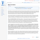
Bile formation is an osmotic secretory process that is driven by the active concentration of bile salts in the bile canaliculi. Bile acids are produced from cholesterol and prior to being excreted from hepatocytes are bound to specific amino acids allowing them to exist as bile salts. One side of the bile salt molecule is negatively charged (hydrophilic) whilst the other is hydrophobic allowing bile salts to form micelles once a certain bile salt concentration has been reached.

Subject:
Life Science
Anatomy/Physiology
Material Type:
Diagram/Illustration
Reading
Provider:
WikiVet
Provider Set:
Anatomy & Physiology
Date Added:
02/05/2015
Blastocyst Embryonic Development - Anatomy & Physiology
Only Sharing Permitted
CC BY-NC-ND
Rating
0.0 stars

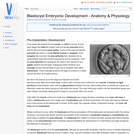
Once sperm has entered the the oocyte, an ootid is formed. During early stages the ootid will contain male and female pronuclei along with the first and second polar bodies. Fusion of the male and female pronuclei will result in a single diploid nucleus or syngamy. Once syngamy has occurred, the zona pellucida then develops into an imprenetrable layer that prevents polyspermy and so polyploidy. Once the zona pellucida has developed, the ootid is now referred to as a zygote (diploid) and will begin undergoing mitotic divisions via a cleavage process that will begin to give rise to daughter cells called blastomeres. These cleavage divisions will begin to produce a 4-celled embryo and then an 8-celled embryo.

Subject:
Life Science
Anatomy/Physiology
Material Type:
Diagram/Illustration
Reading
Provider:
WikiVet
Provider Set:
Anatomy & Physiology
Date Added:
02/05/2015
Blood Brain Barrier - Anatomy & Physiology
Only Sharing Permitted
CC BY-NC-ND
Rating
0.0 stars

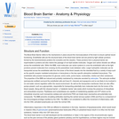
The Blood Brain Barrier refers to the mechanisms in place around the microvasculature of the brain to ensure optimal neural functioning. Endothelial cells are the structural basis of the blood brain barrier and are joined by tight cellular junctions formed by the transmembrane proteins the occludins and the claudins.

Subject:
Life Science
Anatomy/Physiology
Material Type:
Diagram/Illustration
Reading
Provider:
WikiVet
Provider Set:
Anatomy & Physiology
Date Added:
02/05/2015
Blood Cells - Overview
Only Sharing Permitted
CC BY-NC-ND
Rating
0.0 stars

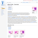
Blood cells develop in the bone marrow from a common stem cell in the process known as haematopoiesis. Once mature, cells are divided into groups that reflect their morphological and functional characteristics including the erythrocytes, or red blood cells, the granulocytes, the agranulocytes and the megakaryocytes.

Subject:
Life Science
Material Type:
Diagram/Illustration
Reading
Provider:
WikiVet
Provider Set:
Blood
Date Added:
02/11/2015
Blood Pressure
Only Sharing Permitted
CC BY-NC-ND
Rating
0.0 stars

This page has links to many topics centered around blood pressure: blood pressure measurement, physiology, kidney control of blood pressure, renal blood pressure, and the renin angiotensin aldosterine system

Subject:
Life Science
Anatomy/Physiology
Material Type:
Diagram/Illustration
Reading
Provider:
WikiVet
Provider Set:
Anatomy & Physiology
Date Added:
02/05/2015
Bone & Cartilage Development - Anatomy & Physiology
Only Sharing Permitted
CC BY-NC-ND
Rating
0.0 stars

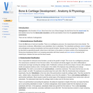
Osteogenesis is the formation of bone. Bone forms from one of three lineages; the skull forms from the neural crest; the limb skeleton forms from the lateral plate mesoderm; and the axial skeleton forms from the paraxial mesoderm (sclerotome).

Subject:
Life Science
Anatomy/Physiology
Material Type:
Diagram/Illustration
Reading
Provider:
WikiVet
Provider Set:
Anatomy & Physiology
Date Added:
02/05/2015
Bovine Anaesthesia
Only Sharing Permitted
CC BY-NC-ND
Rating
0.0 stars

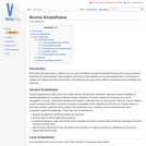
Due to their size and anatomy, cattle are not very good candidates for general anaesthesia meaning that many procedures performed use local techniques. Most surgeries performed on these patients can be done standing with a small amount of sedation and regional and local nerve blocks. Local techniques are also used in cattle for procedures such as castration and dehorning. General anaesthesia is rarely carried out in these species.

Subject:
Life Science
Material Type:
Diagram/Illustration
Reading
Provider:
WikiVet
Provider Set:
Anasthesia
Date Added:
02/27/2015
Bronchi and Bronchioles - Anatomy & Physiology
Only Sharing Permitted
CC BY-NC-ND
Rating
0.0 stars

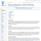
The trachea bifurcates at the levels of the 4th-6th intercostal space, approximately halfway between the thoracic inlet and the diaphragm. It divides into two principle bronchi, tubes which conduct air into the lungs, and they divide into two lobar bronchi for the left lung, and into four lobar bronchi for the right lung. These further divide into smaller bronchi and bronchioles within the lung tissue.

Subject:
Life Science
Anatomy/Physiology
Material Type:
Diagram/Illustration
Reading
Provider:
WikiVet
Provider Set:
Anatomy & Physiology
Date Added:
02/05/2015
Bursa of Fabricius
Only Sharing Permitted
CC BY-NC-ND
Rating
0.0 stars

The Bursa of Fabricus is a primary lymphoid organ found in birds. The bursa was the first place that a certain subset of lymphocytes was observed and consequently they were named B lymphocytes (bursa of Fabricius or bursa equivalent organs). The bursa is involved in the differentiation of B lymphocytes.

Subject:
Life Science
Anatomy/Physiology
Material Type:
Diagram/Illustration
Reading
Provider:
WikiVet
Provider Set:
Anatomy & Physiology
Date Added:
02/05/2015
CNS Development - Anatomy & Physiology
Only Sharing Permitted
CC BY-NC-ND
Rating
0.0 stars

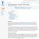
Development of the Central Nervous System (CNS) includes development of the brain, spinal cord, optic and auditory systems, as well as surrounding supporting cells including ependymal cells, astrocytes, oligodendrocytes and microglia. Information within this page will exclude development of the Peripheral Nervous System (PNS) which includes nerve and ganglia formation.

Subject:
Life Science
Anatomy/Physiology
Material Type:
Diagram/Illustration
Reading
Provider:
WikiVet
Provider Set:
Anatomy & Physiology
Date Added:
02/05/2015
CNS Vasculature - Anatomy & Physiology
Only Sharing Permitted
CC BY-NC-ND
Rating
0.0 stars

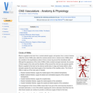
Blood is supplied to the brain from a ventral arterial supply in all species; from a circle of arteries called the Circle of Willis (also called the cerebral arterial circle or arterial circle of Willis) which lies ventrally to the hypothalamus where it forms a loose ring around the infundibular stalk. Although the appearance of the circle of Willis is fairly constant amongst mammals, the sources of blood supply to the circle and the direction of flow around the circle are very species specific. Blood is supplied to the brain by the internal carotid artery in dogs and horses whilst in other domestic species the main blood supply is from branches of the maxillary artery.

Subject:
Life Science
Anatomy/Physiology
Material Type:
Diagram/Illustration
Reading
Provider:
WikiVet
Provider Set:
Anatomy & Physiology
Date Added:
02/05/2015
Caecum - Anatomy & Physiology
Only Sharing Permitted
CC BY-NC-ND
Rating
0.0 stars

The cecum is a blind ending diverticulum of the large intestine and it exists at the junction of the ileum and the ascending colon. Its size and physiological importance varies between species. It is a site of microbial fermentation, absorption and transportation.

Subject:
Life Science
Anatomy/Physiology
Material Type:
Diagram/Illustration
Reading
Provider:
WikiVet
Provider Set:
Anatomy & Physiology
Date Added:
02/05/2015
Camelid Anaesthesia
Only Sharing Permitted
CC BY-NC-ND
Rating
0.0 stars

Camelids are becoming more common in general practice and so an understanding of anaesthesia techniques is becoming more important. The same techniques used in other species can be adapted and used in camelids including both local and general anaesthesia.

Subject:
Life Science
Material Type:
Diagram/Illustration
Reading
Provider:
WikiVet
Provider Set:
Anasthesia
Date Added:
02/27/2015
Camelid Stomach - Anatomy & Physiology
Only Sharing Permitted
CC BY-NC-ND
Rating
0.0 stars

Camelids have a similar digestive structure to other ruminants, although camelids only have three separate stomach compartments compared to the four found in domestic species. The first element of the camelid GI tract, known as C1, can be compared to the rumen whilst the second, known as C2 can be compared to the reticulum. The final element of the tract, C3 can be compared to the abomasum. Therefore camelids do not have a structure comparable to an omasum.

Subject:
Life Science
Anatomy/Physiology
Material Type:
Diagram/Illustration
Reading
Provider:
WikiVet
Provider Set:
Anatomy & Physiology
Date Added:
02/05/2015
Cardiorespiratory System Overview - Anatomy & Physiology
Only Sharing Permitted
CC BY-NC-ND
Rating
0.0 stars

The mammalian cardiovascular and respiratory systems have evolved primarily to provide the tissues of the body with oxygen and to remove carbon dioxide. The cardiorespiratory system also has metabolic and heat exchange roles.

Subject:
Life Science
Anatomy/Physiology
Material Type:
Diagram/Illustration
Reading
Provider:
WikiVet
Provider Set:
Anatomy & Physiology
Date Added:
02/05/2015
Care plans
Only Sharing Permitted
CC BY-NC-ND
Rating
0.0 stars

Written nursing care plans ensure that the nurse responsible for patient care at any time during the animal's stay in the practice is confident to manage and treat the patient, to talk to the owners and give accurate updates on their animal's care, and to feel that the best possible care has been given to the animal at all times. Care plans require skill to write and this is something that improves with practise.

Subject:
Health, Medicine and Nursing
Life Science
Material Type:
Diagram/Illustration
Reading
Provider:
WikiVet
Date Added:
02/27/2015
Carnivore Mammary Gland - Anatomy & Physiology
Only Sharing Permitted
CC BY-NC-ND
Rating
0.0 stars

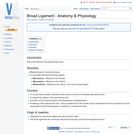
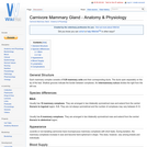
Each mammary complex consists of 5-20 mammary units and their corresponding ducts. The ducts open separately on the tip of the teat. Shallow grooves indicate the border between complexes. An intermammary sulcus divides the right from the left row.

Subject:
Life Science
Anatomy/Physiology
Material Type:
Diagram/Illustration
Reading
Provider:
WikiVet
Provider Set:
Anatomy & Physiology
Date Added:
02/05/2015
Case Study: Is it Time for an NPIP like Program for the US Pork Industry?
Only Sharing Permitted
CC BY-NC-ND
Rating
0.0 stars

The National Poultry Improvement Plan (NPIP) is an industry, state, and federal partnership that has long played a central role in bettering the health of US poultry and improving the competitiveness of the US poultry and egg industries.[1] The objective of this communication is to report the findings of a case study of the NPIP undertaken in 2018. The primary aims of the study included seeking a more in-depth understanding of the NPIP, clarifying how NPIP differs from and complements the Secure Food Supply Plans for the US poultry and egg industries, and assessing the needs and potential applications for establishing a similar program for the US pork industry (e.g., “US Swine Health Improvement Plan”).

Subject:
Agriculture
Nutrition
Zoology
Material Type:
Case Study
Provider:
Iowa State University
Author:
James Roth
Jeffrey Zimmerman
Kerry Leedom-Larson
Pamela Zaabel
Rodger Main
Date Added:
03/09/2020