Hair germs begin from an aggregation of keratinocytes in the stratum basale …
Hair germs begin from an aggregation of keratinocytes in the stratum basale of the epidermis. The initiating factor is the underlying dermal fibroblast cells. The keratinocytes elongate, divide and relocate to the dermis. Dermal fibroblasts then form a dermal papilla beneath the hair germ. This causes stimulation of the basal stem cells to up-regulate their cycle, producing cells that will keratinise and form the hair shaft. Two swellings form on the shaft, one containing stem cells for follicle regeneration, the other becomes a sebaceous gland which will secrete sebum onto the hair shaft. The follicles develop from an ectodermal bud which invades the mesenchyme during embryonic development. The mesoderm also condenses during the development creating an outer mesodermal component to the embedded part of the hair.
Hair germs begin from an aggregation of keratinocytes in the stratum basale …
Hair germs begin from an aggregation of keratinocytes in the stratum basale of the epidermis. The initiating factor is the underlying dermal fibroblast cells. The keratinocytes elongate, divide and relocate to the dermis. Dermal fibroblasts then form a dermal papilla beneath the hair germ. This causes stimulation of the basal stem cells to up-regulate their cycle, producing cells that will keratinise and form the hair shaft.
The formation of the mammalian heart is a fairly complex process. It …
The formation of the mammalian heart is a fairly complex process. It begins when angiogenic mesodermal cells in the cardiogenic plate coalesce to form the endocardial tubes. The endocardial tubes then fuse to form a single duct, the cardiac tube. This undergoes a process of distension, folding and septation and a four chambered, dual circuit pump is formed . The simple heart seen in fish or amphibians forms via the same path but development ceases at an earlier stage.
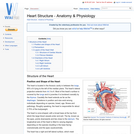
The heart is located in the thoracic cavity in between the lungs, …
The heart is located in the thoracic cavity in between the lungs, 60% of it lying to the left of the median plane. The hearts lateral projection extends from rib 3 to 6. Most of the hearts surface is covered by the lungs and in juveniles it is bordered cranially by the thymus. Caudally the heart extends as far as the diaphragm. Variations in position and size exist among individuals depending on species, breed, age, fitness and pathology. Roughly speaking, the heart is responsible for about 0.75% of the bodyweight.
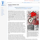
Hepatic stellate cells (HSC) can also be referred to as vitamin A-storing …
Hepatic stellate cells (HSC) can also be referred to as vitamin A-storing cells, lipocytes, interstitial cells, fat-storing cells and Ito cells. HSC exist in the space between parenchymal cells and sinusoidal endothelial cells of the hepatic lobule and store 80% of retinoids in the whole body as retinyl palmitate in lipid droplets in the cytoplasm. In physiological conditions, these cells play pivotal roles in the regulation of retinoid homeostasis; they express specific receptors for retinol-binding protein (RBP), a binding protein specific for retinol, on their cell surface, and take up the complex of retinol and RBP by receptor-mediated endocytosis.
Heterophils are the most abundant granulocyte in most avian species and occur …
Heterophils are the most abundant granulocyte in most avian species and occur alongside lymphocytes, monocytes, eosinophils and basophils in avian blood. These cells are also found in some reptile and mammalian species.
The hind brain is also called the rhombencephalon and is the brain …
The hind brain is also called the rhombencephalon and is the brain stem that provides the connection between the spinal cord and the rest of the brain. The hind brain contains many vital structures including the Medulla Oblongata, the Pons (the link between the cerebellum, forebrain and mid-brain) and the majority of the cranial nerves, III to XII. In general the brain stem governs essential functions that are carried out sub-consciously via reflexes.
Hindgut fermenters are evolved to eat a herbivorous diet. Such a diet …
Hindgut fermenters are evolved to eat a herbivorous diet. Such a diet includes large quantities of insoluble plant carbohydrates, such as cellulose. Mammals cannot digest these insoluble carbohydrates as they lack the essential enzymes, such as cellulase. However it is important that they do digest these carbohydrates as there is insufficient quantity of soluble carbohydrates in plant material. Some microbes do have the enzymes to digest these insoluble carbohydrates and so hindgut fermenters hold a symbiotic relationship with these microbes. Hindgut fermenters have anatomical adaptations to allow for an expanded microbial population. The products of fermentation are volatile fatty acids. It is important to supply a source of fibre in their diet as it stimulates peristalsis in the gut and prevents a build up of gas.
The hoof is defined from a physiologic perspective as the modified skin …
The hoof is defined from a physiologic perspective as the modified skin covering the tip of the digit and all enclosed structures. The hoof provides protection to the distal limb and is formed by keratinisation of the epithelial layer and modification of the underlying dermis. The keratin in the epidermis, when thickened and cornified, is referred to as horn. Horn makes up the outer surface if the hoof and is particularly resistant to mechanical and chemical damage.
The keratin in the epidermis, when cornified and thickened, is referred to …
The keratin in the epidermis, when cornified and thickened, is referred to as horn. Horn is particulary resistant to mechanical and chemical damage. The dermis of horn gives the structures their 3-D structure and shape. Cattle, some sheep, goats and antelope posess horns and these are permanent organs. Breeds without horns are termed polled breeds. Deer posess antlers, which are temporary organs that develop during the rutting season and are then shed.
Lysozyme is one of the major bactericidal agents in secretions and particularly …
Lysozyme is one of the major bactericidal agents in secretions and particularly helps to protect vulnerable sites such as the eyes and nasal passages. The lysoszyme exerts bactericidal effects by digesting bacterial cell walls. The complement system is a group of about 30 proteins within the body fluids of all vertebrates and some invertebrates. The main functions of complement are to promote phagocytosis or causes lysis of an invading organism.
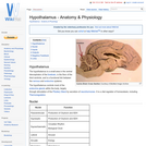
The hypothalamus is a small area in the ventral diencephalon of the …
The hypothalamus is a small area in the ventral diencephalon of the forebrain, in the floor of the third ventricle, and is a functional link between the nervous and endocrine systems.
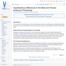
The hypothalamus is inherently female. Testosterone 'defeminizes' the brain during embryogenesis and …
The hypothalamus is inherently female. Testosterone 'defeminizes' the brain during embryogenesis and eliminates the GnRH surge centre in males. The female foetus has no testes to produce testosterone, thus developes a hypothalamic GnRH surge centre.
Hypothesis tests are very commonly used in epidemiological investigations, and a wide …
Hypothesis tests are very commonly used in epidemiological investigations, and a wide number of tests are available. These can be classified into groups according to the data types in question, according to whether a specific underlying distribution is assumed when performing the test (in which case, the test is known as a parametric test), and according to whether or not the data are matched or independent (i.e. whether comparisons are being made at the individual level or the group level). As described earlier, qualitative data are not numerical in nature, and include categorical and ordinal data (such as the breed of dog, or the body condition score of a cow). Quantitative data are numerical, and include variables such as weight, age and height.
The ileum is the terminal portion of the small intestine and continues …
The ileum is the terminal portion of the small intestine and continues from the jejunum. It opens into the caecum at the ileocaecal orifice. The intestinal epithelium is mainly absorptive, with much less digestion occurring compared to the duodenum and the jejunum.
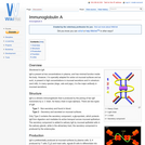
IgA is present at low concentrations in plasma, and has minimal function …
IgA is present at low concentrations in plasma, and has minimal function inside the body. However, it is specially adapted for action at mucosal surfaces and as such, is present in high concentrations in mucosal secretions and in colostrum (and milk). In many species (dogs, cats and pigs), it is the major antibody in
IgD is present in ruminants, pigs, dogs and rodents but has not …
IgD is present in ruminants, pigs, dogs and rodents but has not been identified in horses, cats, rabbits and chickens. It is mainly expressed on the surface of B-cells i.e. it is never secreted.
Unlike IgM, IgG and IgA, IgE does not function as a soluble …
Unlike IgM, IgG and IgA, IgE does not function as a soluble antibody, with binding to Fc? receptors required before it can bind to the target antigen, and is found in low levels in blood plasma. Like IgA, it is produced by plasma cells and is mainly localised to mucosal surfaces.
IgG is the major antibody in blood plasma, and constitutes at least …
IgG is the major antibody in blood plasma, and constitutes at least 80% of all antibodies in the body. It is the smallest immunoglobulin, so can readily leave the blood plasma and enter tissues. They can also cross the placenta, providing adaptive immunity to the foetus when the mother is under attack. IgG is also present in breast milk.
IgM is the primordial antibody and, although a monomer, is secreted as …
IgM is the primordial antibody and, although a monomer, is secreted as a pentamer (five monomers joined by disulphide bonds with two monomers joined by a J chain). This gives it ten identical antigen binding sites although IgM usually has relatively low affinity for its antigen. Its heavy chain is type mu (ľ).
No restrictions on your remixing, redistributing, or making derivative works. Give credit to the author, as required.
Your remixing, redistributing, or making derivatives works comes with some restrictions, including how it is shared.
Your redistributing comes with some restrictions. Do not remix or make derivative works.
Most restrictive license type. Prohibits most uses, sharing, and any changes.
Copyrighted materials, available under Fair Use and the TEACH Act for US-based educators, or other custom arrangements. Go to the resource provider to see their individual restrictions.