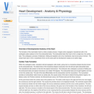
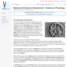
The formation of the mammalian heart is a fairly complex process. It …
The formation of the mammalian heart is a fairly complex process. It begins when angiogenic mesodermal cells in the cardiogenic plate coalesce to form the endocardial tubes. The endocardial tubes then fuse to form a single duct, the cardiac tube. This undergoes a process of distension, folding and septation and a four chambered, dual circuit pump is formed . The simple heart seen in fish or amphibians forms via the same path but development ceases at an earlier stage.
The hindlimb deep veins are very closely related to their respective arteries. …
The hindlimb deep veins are very closely related to their respective arteries. Essentially the lay out of the veins is similar in all domestic species.
Olfaction is the sense of smell, which is the ability to perceive …
Olfaction is the sense of smell, which is the ability to perceive and distinguish odours. Most mammals have a good sense of smell, but most birds generally do not. The sense of smell is well-developed in carnivores (predators) and ungulates (prey). Fish also have a fairly well-developed sense of smell. Olfactory and gustatory receptors can combine to contribute to flavour.
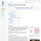
The pharynx is part of both the respiratory and digestive system. Both …
The pharynx is part of both the respiratory and digestive system. Both systems have entrances to the pharynx but they are separated from each other by the soft palate. During exercise or during respiratory distress, the mouth can be used as an additional opening of the respiratory system and then the oropharynx also becomes an air-way.
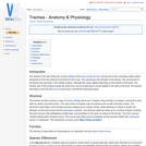
The trachea is the tube linking the cricoid cartilage of the larynx …
The trachea is the tube linking the cricoid cartilage of the larynx to the bronchi, forming part of the conducting system which transports air from the external environment to the lungs. The oesophagus lies dorsally to the trachea. The cervical part of the trachea lies generally in the median position, although this varies slightly depending on the position of the head. The thoracic part of the trachea crosses the aortic arch, thus its positioning is moved slightly to the right at this level. The trachea bifurcates to form the two bronchi at the level of the 4th-6th intercostal space.
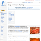
The lungs are the site for gaseous exchange, and are situated within …
The lungs are the site for gaseous exchange, and are situated within the thoracic cavity. They occupy approximately 5% of the body volume in mammals when relaxed, and their elastic nature allows them to expand and contract with the processes of inspiration and expiration.
Once sperm has entered the the oocyte, an ootid is formed. During …
Once sperm has entered the the oocyte, an ootid is formed. During early stages the ootid will contain male and female pronuclei along with the first and second polar bodies. Fusion of the male and female pronuclei will result in a single diploid nucleus or syngamy. Once syngamy has occurred, the zona pellucida then develops into an imprenetrable layer that prevents polyspermy and so polyploidy. Once the zona pellucida has developed, the ootid is now referred to as a zygote (diploid) and will begin undergoing mitotic divisions via a cleavage process that will begin to give rise to daughter cells called blastomeres. These cleavage divisions will begin to produce a 4-celled embryo and then an 8-celled embryo.
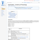
Gastrulation is the process of forming the three germ layers; ectoderm, mesoderm …
Gastrulation is the process of forming the three germ layers; ectoderm, mesoderm and endoderm. It is achieved through a series of highly coordinated cell movements. Cells that will form the endodermal and mesodermal organs are brought inside the embryo, whilst cells that will form ectoderm move to spread out over the outside of the embryo.
The intermediate mesoderm exists as a strip of tissue between the lateral …
The intermediate mesoderm exists as a strip of tissue between the lateral plate mesoderm and somites. It gives rise to the urinary system and some parts of the reproductive system. Kidney development includes three forms: Pronephros, Mesonephros, and Metanephros. Mammals develop all three, and continue to use the metanephros in adult life. More primitive animals have only the first one or two.
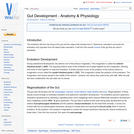
The endoderm will form the lining of the gut and the organs …
The endoderm will form the lining of the gut and the organs that develop from it. Splanchnic mesoderm surrounds the endoderm and orginates from the lateral plate mesoderm. It will form the smooth muscle of the gut that are used in peristalsis.
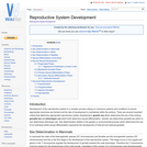
Development of the reproductive system is a complex process relying on numerous …
Development of the reproductive system is a complex process relying on numerous systems and conditions to ensure appropriate structures are formed and the rate of development is maintained within the embryo. There are several important criteria that determine appropriate reproductive system development; genetic sex which determines the sex of the embryo, gonadal sex and phenotypic sex which both determine sexual differentiation. Genetic sex determines gonadal sex which in turn determines phenotypic sex. Sex determination relates to the genetic or environmental process which determines the sex of an individual whilst sexual differentiation represents the development of internal and external genitalia.
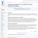
The hypothalamus is inherently female. Testosterone 'defeminizes' the brain during embryogenesis and …
The hypothalamus is inherently female. Testosterone 'defeminizes' the brain during embryogenesis and eliminates the GnRH surge centre in males. The female foetus has no testes to produce testosterone, thus developes a hypothalamic GnRH surge centre.
The pituitary gland, or hypophysis is an elongated appendage of the brain …
The pituitary gland, or hypophysis is an elongated appendage of the brain lying within a bony cavity of the sphenoid bone in the base of the skull - the Sella Turcica. The hypophysis is suspended from the hypothalamus by a thin stalk. It lies between the more rostral optic chiasma, and the more caudal mammillary bodies.
The thymus has a key role in the maturation of prothymocytes into …
The thymus has a key role in the maturation of prothymocytes into mature T cells. In juvenile animals the thymus produces significant numbers of new T lymphocytes but as the animal matures this production decreases and T cell population is maintained by division of mature T cells.
The pituitary gland, or hypophysis is an elongated appendage of the brain …
The pituitary gland, or hypophysis is an elongated appendage of the brain lying within a bony cavity of the sphenoid bone in the base of the skull - the Sella Turcica. The hypophysis is suspended from the hypothalamus by a thin stalk. It lies between the more rostral optic chiasma, and the more caudal mammillary bodies.
The forebrain (proencephalon) is the largest part of the brain, most of …
The forebrain (proencephalon) is the largest part of the brain, most of which is cerebrum. Other important structures found in the forebrain include the thalamus , the hypothalamus and the limbic system. The cerebrum is divided into two cerebral hemispheres connected by a mass of white matter known as the corpus callosum.
Nerves of the peripheral nervous system (PNS) are composed of numerous bundles …
Nerves of the peripheral nervous system (PNS) are composed of numerous bundles of nerve fibers that are surrounded by connective tissue. This connective tissue also contains a specific layer that is specialised to neurons; the peri-neurium. The outer layer of this connective tissue is called the epineurium and it surrounds both the perineurium and the nerve itself. Individual neurons found within each bundle are surrounded by the endoneurium.
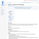
The Oviduct is the tube that links the ovary to the uterus …
The Oviduct is the tube that links the ovary to the uterus and which the ovulated oocyte travels down to become fertilised by sperm present in the female tract. It is also refered to as the Fallopian tube, Uterine tube or Ovarian tube.
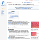
Luteinisation occurs after ovulation and the collapse of the follicle. The number …
Luteinisation occurs after ovulation and the collapse of the follicle. The number of corpora lutea formed in the ovary at any one time is directly proportional to the number of oocytes ovulated. Therefore many corpora lutea will be visible on the ovary of polytocous animals. During Luteinisation there is an increase in both the size and weight due to hyperplasia (increase in cell number) and hypertrophy (increase in cell size) within the developing corpus luteum.
No restrictions on your remixing, redistributing, or making derivative works. Give credit to the author, as required.
Your remixing, redistributing, or making derivatives works comes with some restrictions, including how it is shared.
Your redistributing comes with some restrictions. Do not remix or make derivative works.
Most restrictive license type. Prohibits most uses, sharing, and any changes.
Copyrighted materials, available under Fair Use and the TEACH Act for US-based educators, or other custom arrangements. Go to the resource provider to see their individual restrictions.