(This case study was added to OER Commons as one of a …
(This case study was added to OER Commons as one of a batch of over 700. It has relevant information which may include medical imagery, lab results, and history where relevant. A link to the final diagnosis can be found at the end of the case study for review. The first paragraph of the case study -- typically, but not always the clinical presentation -- is provided below.)
A 55-year-old woman presented with bilateral hip and rib pain. A chest radiograph revealed multiple bilateral rib fractures with callus formation. Insufficiency fractures of the right superior and inferior pubic rami and ischium and possibly of the sacrum were noted on hip and pelvic radiographs, and a subsequent MRI showed avascular necrosis of the left femoral head. Laboratory studies demonstrating hypophosphatemia, in combination with the patient's clinical presentation of osteomalacia, prompted further investigation for the underlying cause.
(This case study was added to OER Commons as one of a …
(This case study was added to OER Commons as one of a batch of over 700. It has relevant information which may include medical imagery, lab results, and history where relevant. A link to the final diagnosis can be found at the end of the case study for review. The first paragraph of the case study -- typically, but not always the clinical presentation -- is provided below.)
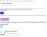
A term female neonate presented for excision of two prenatally detected oral cavity masses. The pregnancy was uncomplicated, and the neonate was delivered via Caesarean section without complications. Immediately following delivery, the patient was transferred to the operating room. Intra-operative findings included a 1 cm mass of the ventral tongue and a 4 cm mass of the left maxillary alveolar ridge, both of which were excised. At time of discharge, she was feeding orally without issue and was stable on room air.
(This case study was added to OER Commons as one of a …
(This case study was added to OER Commons as one of a batch of over 700. It has relevant information which may include medical imagery, lab results, and history where relevant. A link to the final diagnosis can be found at the end of the case study for review. The first paragraph of the case study -- typically, but not always the clinical presentation -- is provided below.)
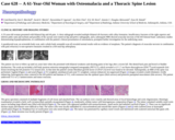
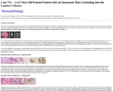
A 72 year old female with no significant past medical history who was under treatment and follow up for her chronic low back pain was referred to our center for evaluation of an incidentally discovered intramedullary lesion in her thoracic spine after her recent fall. She didn't have any neurologic deficits at the time, including weakness or any bladder or bowl dysfunction and on exam she was neurologically intact. On her further work up, MRI of thoracic spine revealed a well-defined intramedullary lesion with an intradural component measuring about 1 cm at the level of T10. (Fig 1, left) There was a mildly increased T2 signal with enhancement after gadolinium administration. Chronic superior endplate compression fracture of T9 and L1 vertebral body was also noted. The intramedullary lesion was considered an incidental finding unrelated to her symptoms therefore the initial plan was close observation, given her unwillingness for any surgical intervention, in addition to the small size of the lesion, lack of neurological deficits and its benign appearance on imaging. Based on imaging, the differential was a low-grade astrocytoma, ependymoma or an intramedullary schwannoma. After about 3 years, in a follow up visit, we noticed a mild weakness in her left lower extremity and some gait unsteadiness. This was accompanied by a significant increase in the size of the lesion (Fig 1, right), so she agreed to an excisional biopsy. Intraoperatively the lesion was noted to be a bluish, firm mass with a good plane at the level of T10/11.
(This case study was added to OER Commons as one of a …
(This case study was added to OER Commons as one of a batch of over 700. It has relevant information which may include medical imagery, lab results, and history where relevant. A link to the final diagnosis can be found at the end of the case study for review. The first paragraph of the case study -- typically, but not always the clinical presentation -- is provided below.)
A 47-year old Hispanic male with end-stage AIDS was admitted to the hospital with pneumonia, altered mental status and right-sided weakness. His previous medical history was obscure. He had never received antiretroviral treatment. Upon his arrival he was cachectic, tachycardic and lethargic. His neurologic exam showed left facial palsy, right hemiparesis, hyperreflexia, and a positive right Babinski sign. He had no signs of meningeal irritation. On admission his white blood cell count was 8400/mm3 (98% neutrophils), and his CD4+ cell count was 8 cells/mm3. The serum cryptococcal antigen was negative. IgG and IgM toxoplasma antibodies were positive. A MRI was ordered which was suggestive of neurotoxoplasmosis. He was started on piperacillin-tazobactam, clarithromycin and trimethoprim / sulfamethoxazole for pneumonia, valgancyclovir for skin biopsy proven CMV infection, fluconazol for oral candidiasis and pyrimethamine and clindamycin for toxoplasmosis. Although he continued disoriented and sleepy, he was stable for a few days. On the 18th hospital day he developed severe respiratory distress. He became stuporous and presented right midriasis and ptosis, impaired adduction of the right eye, and generalized seizures. He required immediate ventilatory support and high doses of vasoactive amines. Intravenous amphothericin B infusion was started in order to broaden the antimicrobial coverage, but the patient died five days later.
(This case study was added to OER Commons as one of a …
(This case study was added to OER Commons as one of a batch of over 700. It has relevant information which may include medical imagery, lab results, and history where relevant. A link to the final diagnosis can be found at the end of the case study for review. The first paragraph of the case study -- typically, but not always the clinical presentation -- is provided below.)
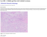
We present the case of a 61-year-old male with a longstanding history of pituitary macro-prolactinoma medically treated with high dose cabergoline without significant reduction in tumor size. The pituitary lesion was first discovered more than 11 years prior to presentation and had been routinely monitored since that time with serial magnetic resonance imaging (MRI) scans and hormone profile testing. Pre-operative MRI in July 2014 revealed an enlarged, remodeled sella and a pituitary macroadenoma measuring approximately 2 cm in greatest dimension (Figure 1). Pre-operative laboratory values from March 2015 showed no evidence for an active neuroendocrine tumor. Detailed serology included a prolactin level of 7.6 ng/mL (reference range, 2.1 - 17.7 ng/mL) and a growth hormone level of 0.07 ng/mL (reference range, 0.01 - 0.97 ng/mL). Cortisol, adrenocorticotropic, luteinizing, and follicle stimulating hormones were also within normal serologic range. The patient underwent endoscopic endonasal resection of the pituitary mass, and a gross total resection was achieved. Intraoperatively, the lesion was described as extremely gritty and nodular. Post-operatively, the patient remained asymptomatic for several months despite a slight increase in his prolactin level to 8.9 ng/mL and remains asymptomatic to date almost one year post surgery. His postoperative endocrinologic pituitary hormonal profile is unchanged from his preoperative levels.
(This case study was added to OER Commons as one of a …
(This case study was added to OER Commons as one of a batch of over 700. It has relevant information which may include medical imagery, lab results, and history where relevant. A link to the final diagnosis can be found at the end of the case study for review. The first paragraph of the case study -- typically, but not always the clinical presentation -- is provided below.)
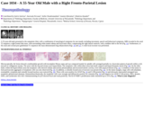
A 44-year-old man with a past medical history of arterial hypertension, hypercholesterolemia, cigarette smoking (45 pack-years) and obesity (BMI 32.8) presented to our department with a 3-month history of right-sided facial numbness. Four weeks prior to admission he experienced a single episode of involuntary muscle movements on the left-side of his body. His neurologic exam was normal and initial laboratory results including CBC and blood chemistry were within normal range. A magnetic resonance imaging (MRI) scan of the patient's brain (Figure 1) showed a 7.3x4.9x3.6 cm, right fronto-parietal, extra-axial space-occupying lesion with lobulated contrast-enhancement and mild perifocal edema. The superior sagittal sinus was slightly compressed and the overlying cranium was infiltrated. The patient underwent angio-embolization of the lesion and two days later a right fronto-temporo-parietal craniectomy was performed. The tumor was resected subtotally, leaving a thin superficial infiltrative layer on eloquent cortex. The infiltrated cranium was reconstructed using polymethyl-methacrylate (PMMA) cranioplasty and the resected dura was replaced by a neuropatch. Postoperatively, an MRI of the spine and a lumbar puncture did not show any evidence for disease dissemination. The patient had no neurological deficit and underwent adjuvant radiation therapy of the tumor bed (36 Gy) and 2-years after diagnosis he was clinically and radiologically disease-free.
(This case study was added to OER Commons as one of a …
(This case study was added to OER Commons as one of a batch of over 700. It has relevant information which may include medical imagery, lab results, and history where relevant. A link to the final diagnosis can be found at the end of the case study for review. The first paragraph of the case study -- typically, but not always the clinical presentation -- is provided below.)
The patient is a teenager boy who was previously diagnosed with ALL in 2001. He had numerous relapses and received a bone marrow transplant in 2002. He subsequently developed grafts vs host disease and his immunosuppression increased. The patient subsequently presented with visual disturbances and headache over a period of two to three weeks in May 2003. Initially, CT showed a large mass in the in the right temporal lobe. All other radiology was negative. A stereotactic brain biopsy was performed.
(This case study was added to OER Commons as one of a …
(This case study was added to OER Commons as one of a batch of over 700. It has relevant information which may include medical imagery, lab results, and history where relevant. A link to the final diagnosis can be found at the end of the case study for review. The first paragraph of the case study -- typically, but not always the clinical presentation -- is provided below.)
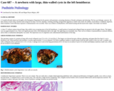
A 49-year-old female, who suffered trauma to the left fronto-parietal region from a gymnasium weight two years previously, presented with a 12-month history of an enlarging lump in the same area. There were no associated visual, sensory or neurological symptoms. On examination there was a large smooth, non-tender, bony hard mass measuring 8 x 6 centimetres at its base, with mild to moderate overlying alopecia. It was non-pulsatile with no bruits. The lesion was percussion dull and there was no regional lymphadenopathy.
(This case study was added to OER Commons as one of a …
(This case study was added to OER Commons as one of a batch of over 700. It has relevant information which may include medical imagery, lab results, and history where relevant. A link to the final diagnosis can be found at the end of the case study for review. The first paragraph of the case study -- typically, but not always the clinical presentation -- is provided below.)
A 62-year old female patient presented with constricted nasal breathing on the right side, disorder of smell and anosmia for the past six months. She had no epistaxis, rhinorrhea or loss of sensitivity in the face. Three months before presentation she suffered from a cold with swelling of the right eyelid that improved under conservative therapy. Physical examination revealed a glazed, bleeding tumor occupying the complete right nasal cavity and right nasopharynx and extending to the opposite site (Figure 1).
(This case study was added to OER Commons as one of a …
(This case study was added to OER Commons as one of a batch of over 700. It has relevant information which may include medical imagery, lab results, and history where relevant. A link to the final diagnosis can be found at the end of the case study for review. The first paragraph of the case study -- typically, but not always the clinical presentation -- is provided below.)
A 5 month-old male infant was brought to the Emergency Department by his parents with persistent, worsening shortness of breath, tachypnea and wheezing. The boy was lethargic, anorexic. He had a history of poor sleep, fussiness and crying for two days. No fever, diarrhea, vomiting, drooling, foreign-body ingestion or stridor were noted. No asthma, recent illness or antibiotic ingestion, home treatment, recent infectious exposure or immunizations were recorded. There was evidence of dehydration, with limited tearing and decreased urine output. Oral intake was limited.
(This case study was added to OER Commons as one of a …
(This case study was added to OER Commons as one of a batch of over 700. It has relevant information which may include medical imagery, lab results, and history where relevant. A link to the final diagnosis can be found at the end of the case study for review. The first paragraph of the case study -- typically, but not always the clinical presentation -- is provided below.)
A male patient in his 60s with a history of urothelial carcinoma metastatic to the liver, status post radical cystoprostatectomy and chemotherapy. Patient later presented with right renal pelvis tumor. The biopsy showed fragments of high grade urothelial carcinoma with inverted growth pattern, with tumor cells positive for uroplakin II + III, CAM 5.2, and GATA-3 by immunohistochemical analysis. Patient was treated with gemcitabine, carboplatin and Enfortumab but progressed, with evidence of new lung and liver metastases. Oncomine NGS testing was performed to identify potential therapeutic targets.
(This case study was added to OER Commons as one of a …
(This case study was added to OER Commons as one of a batch of over 700. It has relevant information which may include medical imagery, lab results, and history where relevant. A link to the final diagnosis can be found at the end of the case study for review. The first paragraph of the case study -- typically, but not always the clinical presentation -- is provided below.)
A 55-year-old male presented to the outpatient clinic with a combination of neurological symptoms for one month, including movement, speech and behavioral symptoms. MRI revealed in the axial T2 sequence a right frontal lobe mass, with surrounding white matter edema and local mass effect, compressing the right lateral ventricle, with a midline shift to the left (Fig. 1a). Furthermore, in the axial and coronal post gadolinium T1 sequences the mass demonstrated ring enhancement (Figs. 1b and 1c). A wide local excision was performed.
(This case study was added to OER Commons as one of a …
(This case study was added to OER Commons as one of a batch of over 700. It has relevant information which may include medical imagery, lab results, and history where relevant. A link to the final diagnosis can be found at the end of the case study for review. The first paragraph of the case study -- typically, but not always the clinical presentation -- is provided below.)
A 31-year-old, previously healthy woman experienced a new-onset generalized seizure with subsequent right-sided weakness in the 37th week of her pregnancy. History revealed no significant nausea or vomiting (other than that associated with the first trimester of pregnancy) and no prior history of seizures. The patient did, however, report a history of slowly-increasing weakness in her right leg over the past several months. Routine prenatal care had been uneventful and negative for gestational diabetes or hypertension.
(This case study was added to OER Commons as one of a …
(This case study was added to OER Commons as one of a batch of over 700. It has relevant information which may include medical imagery, lab results, and history where relevant. A link to the final diagnosis can be found at the end of the case study for review. The first paragraph of the case study -- typically, but not always the clinical presentation -- is provided below.)
The patient was a 60 year-old woman who had inflammatory ductal carcinoma of the left breast diagnosed on a core biopsy in January 2005. An axillary lymph node was positive for metastatic disease on a concurrent FNA. The tumor was found to be ER-positive, PR-negative, and Her2-Neu weakly positive. Workup for further metastatic disease found multiple lesions in the liver and spine [images 1 and 2] as well as a 5 cm mass in the upper pole of the left kidney [image 3].
(This case study was added to OER Commons as one of a …
(This case study was added to OER Commons as one of a batch of over 700. It has relevant information which may include medical imagery, lab results, and history where relevant. A link to the final diagnosis can be found at the end of the case study for review. The first paragraph of the case study -- typically, but not always the clinical presentation -- is provided below.)
The patient is a preadolescent female who presented with nausea, vomiting, blurred vision, diplopia, gait instability and ataxia. An MRI showed a 22 x 21 x 78 mm enhancing tumor with a necrotic center located at the craniocervical junction expanding the medulla oblongata and the proximal aspect of the spinal cord to the level of T3. Additionally, there was a non-enhancing exophytic component of the tumor abutting the left premedullary cistern (Figure 1). The radiologic impression was an infiltrative, likely astrocytic, craniocervical junction tumor with an exophytic component.
This resource in an activity for sports medicine or health science students …
This resource in an activity for sports medicine or health science students with an understanding of anatomy, physiology, common disorders and injuries to the musckeloskeletal system and techniques of assessing injuries including obtaining medical histories and evaluating techniques. This resource provided a template for students to use when practicing technical skills needed for assessment of a knee injury.
(This case study was added to OER Commons as one of a …
(This case study was added to OER Commons as one of a batch of over 700. It has relevant information which may include medical imagery, lab results, and history where relevant. A link to the final diagnosis can be found at the end of the case study for review. The first paragraph of the case study -- typically, but not always the clinical presentation -- is provided below.)
A man in his 50s was diagnosed with Glioblastoma Mulitforme of the left temporal lobe for which he underwent chemotherapy and external beam radiation therapy. He presented suddenly nine months later with progressive aphasia and right-sided hemiplegia. Following a physical and neurological examination it was noted that the patient again had a mass situated in the left temporal lobe (in the previous site of the surgery) associated with significant compression of the brainstem.
(This case study was added to OER Commons as one of a …
(This case study was added to OER Commons as one of a batch of over 700. It has relevant information which may include medical imagery, lab results, and history where relevant. A link to the final diagnosis can be found at the end of the case study for review. The first paragraph of the case study -- typically, but not always the clinical presentation -- is provided below.)
A 21-year old male with a history of intravenous heroin abuse presented to Presbyterian Hospital status post cardiac arrest. The patient was found unconscious by his father with snoring respirations. His father turned to call 911. When he returned from that phone call, he found that his son had stopped breathing. He was found with Seroquel (quetiapine) packets in his room. When paramedics arrived the patient was in asystole.
(This case study was added to OER Commons as one of a …
(This case study was added to OER Commons as one of a batch of over 700. It has relevant information which may include medical imagery, lab results, and history where relevant. A link to the final diagnosis can be found at the end of the case study for review. The first paragraph of the case study -- typically, but not always the clinical presentation -- is provided below.)
A previously healthy, 35-year-old man presented with a four-week history of painful swelling on his right forehead. CT imaging showed a solitary, 1 cm osteolytic lesion within the diploic space of the right frontal bone, involving the inner and outer tables (Figure 1). On MRI, the lesion was heterogeneously enhancing and extended into subgaleal tissue and epidural space. It was resected and the patient was discharged in good condition.
(This case study was added to OER Commons as one of a …
(This case study was added to OER Commons as one of a batch of over 700. It has relevant information which may include medical imagery, lab results, and history where relevant. A link to the final diagnosis can be found at the end of the case study for review. The first paragraph of the case study -- typically, but not always the clinical presentation -- is provided below.)
The patient is a 62-year old man who was suffering from a chronic pyothorax after pneumectomy 44 years ago. In August 2002 a fenestration of the left thoracic wall was performed. Besides several serological signs of inflammation, NSE (neuron-specific enolase) was markedly elevated. Postoperatively, the patient complained of progressive ataxia and a cerebellar biopsy was performed. Despite treatment the patient died from aspergillus sepsis two month later and permission for autopsy was obtained.
No restrictions on your remixing, redistributing, or making derivative works. Give credit to the author, as required.
Your remixing, redistributing, or making derivatives works comes with some restrictions, including how it is shared.
Your redistributing comes with some restrictions. Do not remix or make derivative works.
Most restrictive license type. Prohibits most uses, sharing, and any changes.
Copyrighted materials, available under Fair Use and the TEACH Act for US-based educators, or other custom arrangements. Go to the resource provider to see their individual restrictions.