(This case study was added to OER Commons as one of a …
(This case study was added to OER Commons as one of a batch of over 700. It has relevant information which may include medical imagery, lab results, and history where relevant. A link to the final diagnosis can be found at the end of the case study for review. The first paragraph of the case study -- typically, but not always the clinical presentation -- is provided below.)
The patient was a man in his 60s with new detection of hypertension and chronic kidney disease presenting with nephrotic range proteinuria, and peripheral edema. The patient was a former smoker (50 pack years) and had used NSAID (Celebrex and Ibuprofen) for many years for his chronic back pain, but discontinued 3 months prior to kidney biopsy after he was found to have an elevated serum creatinine(Table 1) and additional pertinent labs (Table 2) include:
(This case study was added to OER Commons as one of a …
(This case study was added to OER Commons as one of a batch of over 700. It has relevant information which may include medical imagery, lab results, and history where relevant. A link to the final diagnosis can be found at the end of the case study for review. The first paragraph of the case study -- typically, but not always the clinical presentation -- is provided below.)
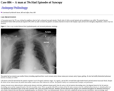
A Caucasian man in his 70's was visiting his neighbors when he had a witnessed syncopal episode. Shortly after, he had a second episode and an ambulance was called. The patient became unresponsive before EMS arrived and required CPR. EMS was able to restore the patient's circulatory function en route to the hospital. Upon arrival in the emergency department, a chest x-ray was obtained.
(This case study was added to OER Commons as one of a …
(This case study was added to OER Commons as one of a batch of over 700. It has relevant information which may include medical imagery, lab results, and history where relevant. A link to the final diagnosis can be found at the end of the case study for review. The first paragraph of the case study -- typically, but not always the clinical presentation -- is provided below.)
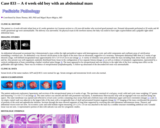
The patient is a 4 week-old male infant born at 41 weeks gestation via Cesarean section to a 26 year-old mother who received good prenatal care. Prenatal ultrasounds performed at 20 weeks and 40 weeks gestational age were unremarkable. The delivery was uneventful. On physical exam in the newborn nursery the baby was noted to have right cryptorchidism and a palpable right-sided abdominal mass.
(This case study was added to OER Commons as one of a …
(This case study was added to OER Commons as one of a batch of over 700. It has relevant information which may include medical imagery, lab results, and history where relevant. A link to the final diagnosis can be found at the end of the case study for review. The first paragraph of the case study -- typically, but not always the clinical presentation -- is provided below.)
The patient was an elderly man with history of chronic lymphocytic leukemia/small lymphocytic lymphoma (CLL/SLL), recently started on ibrutinib, who presented with high fever, chills/rigors, night sweats and altered mental status. He was also noted to be hypotensive, which responded to IV NS. His spleen was enlarged, measuring up to 14.6 cm on ultrasound. He had no known significant recent exposures or travel. This occurred in the winter.
(This case study was added to OER Commons as one of a …
(This case study was added to OER Commons as one of a batch of over 700. It has relevant information which may include medical imagery, lab results, and history where relevant. A link to the final diagnosis can be found at the end of the case study for review. The first paragraph of the case study -- typically, but not always the clinical presentation -- is provided below.)
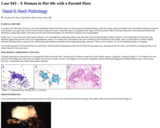
A woman in her 60s with a history of clear cell endometrial adenocarcinoma status post hysterectomy and chemotherapy, and breast cancer status post lumpectomy and radiation therapy, presented to her primary care physician with left eyelid droop. On physical exam, a nontender mass was palpated in the region of the left parotid. MRI of the brain showed no intracranial abnormality but identified an enhancing nodule in the deep portion of the left parotid gland, for which ENT evaluation was recommended.
(This case study was added to OER Commons as one of a …
(This case study was added to OER Commons as one of a batch of over 700. It has relevant information which may include medical imagery, lab results, and history where relevant. A link to the final diagnosis can be found at the end of the case study for review. The first paragraph of the case study -- typically, but not always the clinical presentation -- is provided below.)
MRI revealed an irregular 2.0 x 2.5 x 3.0 cm right frontal lobe ring enhancing mass with edema (Figure 1). The lesion was believed to represent a primary or secondary tumor. A resection of the right frontal lobe lesion revealed a gliotic solid and cystic mass. The resected lesion consisted of irregular tan to pink fragments of tissue measuring 3.5 x 1.2 x 0.5 cm.
(This case study was added to OER Commons as one of a …
(This case study was added to OER Commons as one of a batch of over 700. It has relevant information which may include medical imagery, lab results, and history where relevant. A link to the final diagnosis can be found at the end of the case study for review. The first paragraph of the case study -- typically, but not always the clinical presentation -- is provided below.)
The patient is a 27-year-old male presenting with acute carpopedal spasm, tachypnea and pain in all the extremities. The patient has a history of difficulty speaking. The patient has a history of generalized myalgia and weakness of the body. There is no history of change in his vision or hearing. The patient has no ongoing nausea, vomiting or diarrhea. There are no urinary symptoms and no history of recent head injury. On initial examination, the patient was noted to be alert and oriented and somewhat dysarthric.
(This case study was added to OER Commons as one of a …
(This case study was added to OER Commons as one of a batch of over 700. It has relevant information which may include medical imagery, lab results, and history where relevant. A link to the final diagnosis can be found at the end of the case study for review. The first paragraph of the case study -- typically, but not always the clinical presentation -- is provided below.)
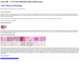
The patient was a 57-year-old gentleman with a 2 week history of nausea, vomiting, abdominal pain and distention who presented with fever, chills, and sharp pain radiating to both groins. The initial CT scan showed a large heterogeneous pelvic soft tissue mass partially encasing bilateral common iliac vessels, rectum, anus, and sigmoid colon with deviation of the urinary bladder but no evidence of colonic obstruction.
(This case study was added to OER Commons as one of a …
(This case study was added to OER Commons as one of a batch of over 700. It has relevant information which may include medical imagery, lab results, and history where relevant. A link to the final diagnosis can be found at the end of the case study for review. The first paragraph of the case study -- typically, but not always the clinical presentation -- is provided below.)
A 19-year-old previously healthy man was admitted in an unconscious state with a conjugate deviation of gaze to the right. Having been intubated and ventilated, he suffered a series of generalized seizures. Cranial MRI showed a slightly enhanced periventricular edema zone in the white matter adjacent to the posterior horn (Fig. 1; A: fluid-attenuated inversion recovery [FLAIR], B: T2-weighed, C: gadolinium enhanced).
(This case study was added to OER Commons as one of a …
(This case study was added to OER Commons as one of a batch of over 700. It has relevant information which may include medical imagery, lab results, and history where relevant. A link to the final diagnosis can be found at the end of the case study for review. The first paragraph of the case study -- typically, but not always the clinical presentation -- is provided below.)
This patient is a 7-month old baby boy who underwent kidney transplantation due to congenital steroid-resistant nephrotic syndrome with progressive renal failure. Additional clinical history included bilateral inguinal exploration followed by right orchidopexy due to cryptorchidism; the left testis had not been found on surgical exploration. Physical examination disclosed a well-developed baby boy with normal male genitalia and a non-palpable left testis.
(This case study was added to OER Commons as one of a …
(This case study was added to OER Commons as one of a batch of over 700. It has relevant information which may include medical imagery, lab results, and history where relevant. A link to the final diagnosis can be found at the end of the case study for review. The first paragraph of the case study -- typically, but not always the clinical presentation -- is provided below.)
An eight year old boy with a history of autism and cerebral palsy was brought to his pediatrician by his mother for new onset staring episodes. According to the mother, the infrequent episodes are typified by the boy stopping suddenly, staring and becoming unresponsive. The mother also noted that the child has been sleeping more in recent weeks. The child's history is negative for convulsions, myoclonic jerks, meningitis, encephalitis or severe head trauma associated with loss of consciousness.
(This case study was added to OER Commons as one of a …
(This case study was added to OER Commons as one of a batch of over 700. It has relevant information which may include medical imagery, lab results, and history where relevant. A link to the final diagnosis can be found at the end of the case study for review. The first paragraph of the case study -- typically, but not always the clinical presentation -- is provided below.)
The patient is a 76 year-old female who is status post partial nephrectomy for renal cell carcinoma, chromophobe subtype, four years prior to presentation. She has had no subsequent recurrence, but now presents with a mass in the anterior medial aspect of her left thigh that had been progressively enlarging over the past nine months. A few weeks before her presentation, the lesion began to grow more rapidly and became painful.
(This case study was added to OER Commons as one of a …
(This case study was added to OER Commons as one of a batch of over 700. It has relevant information which may include medical imagery, lab results, and history where relevant. A link to the final diagnosis can be found at the end of the case study for review. The first paragraph of the case study -- typically, but not always the clinical presentation -- is provided below.)
A 42-year-old man was admitted to the neurosurgery department because of paraparesis and sensory deficits of both feet. The CT scan and MRI examination revealed a solitary intramedullary lesion bulging dorsally from the thoracic spine (T4 level) (Figure 1). Spinal angiography revealed the dense vascularity of the lesion, the presence of feeding and draining vessels, as well as intra-lesional shunting. A gross total resection was performed.
(This case study was added to OER Commons as one of a …
(This case study was added to OER Commons as one of a batch of over 700. It has relevant information which may include medical imagery, lab results, and history where relevant. A link to the final diagnosis can be found at the end of the case study for review. The first paragraph of the case study -- typically, but not always the clinical presentation -- is provided below.)
The patient is an 80-year-old man who presented with the chief complaint of right hemi-scrotal swelling following a history of prostatic adenocarcinoma diagnosed 8 years earlier. The prostate cancer was treated by a combination of radiation therapy and androgen deprivation therapy. Biochemical recurrence of prostatic adenocarcinoma had recently been documented prior to the onset of the swelling. Physical examination revealed a paratesticular mass that was clinically felt to be a benign hydrocele.
(This case study was added to OER Commons as one of a …
(This case study was added to OER Commons as one of a batch of over 700. It has relevant information which may include medical imagery, lab results, and history where relevant. A link to the final diagnosis can be found at the end of the case study for review. The first paragraph of the case study -- typically, but not always the clinical presentation -- is provided below.)
The patient is a woman in her mid-40s who presented to her primary care physician complaining of menorrhagia. In addition to uterine leiomyomas, radiologic work-up revealed a right complex ovarian cystic lesion, measuring approximately 4.5 cm in greatest dimension. The patient had no personal or family history of breast or ovarian cancer. She subsequently underwent a total abdominal hysterectomy and bilateral salpingooopherectomies.
(This case study was added to OER Commons as one of a …
(This case study was added to OER Commons as one of a batch of over 700. It has relevant information which may include medical imagery, lab results, and history where relevant. A link to the final diagnosis can be found at the end of the case study for review. The first paragraph of the case study -- typically, but not always the clinical presentation -- is provided below.)
A 49-year-old right-handed man developed progressive cognitive difficulties over a four month period. Recent memory was impaired. He was unable to do the payroll at his company and would get lost in familiar surroundings. There were word-finding and language difficulties. He had associated fatigue, anorexia, daytime somnolence and weight loss of thirty pounds. Gait imbalance and urinary incontinence developed later.
(This case study was added to OER Commons as one of a …
(This case study was added to OER Commons as one of a batch of over 700. It has relevant information which may include medical imagery, lab results, and history where relevant. A link to the final diagnosis can be found at the end of the case study for review. The first paragraph of the case study -- typically, but not always the clinical presentation -- is provided below.)
A 3-month-old male infant with pain over the left scrotum for two days. The mother noticed bluish discoloration of the left scrotum and took the patient to the hospital. Ultrasound of the scrotum revealed no flow to the left testis and flow to the right testis. Physical examination revealed hard and enlarged left testis, 2 cm, tender to palpation. The right testis and the penis were normal in appearance. Lab test showed normal AFP level.
(This case study was added to OER Commons as one of a …
(This case study was added to OER Commons as one of a batch of over 700. It has relevant information which may include medical imagery, lab results, and history where relevant. A link to the final diagnosis can be found at the end of the case study for review. The first paragraph of the case study -- typically, but not always the clinical presentation -- is provided below.)
A 33-year-old male presented with a 2-week old uncomfortable mass within his left testicle. His serum -hCG was elevated (345.2 mIU/mL). Ultrasound examination demonstrated a heterogeneous, hypoechoic mass. The patient subsequently underwent left radical orchiectomy and epididymectomy. Two weeks after the operation -hCG level was less than 2 mIU/ml. Clinical and radiological staging was negative.
(This case study was added to OER Commons as one of a …
(This case study was added to OER Commons as one of a batch of over 700. It has relevant information which may include medical imagery, lab results, and history where relevant. A link to the final diagnosis can be found at the end of the case study for review. The first paragraph of the case study -- typically, but not always the clinical presentation -- is provided below.)
The patient is a 15-year-old adolescent male with a history of developmental delay who presented at 8 to 10 years of age with loss of peripheral vision and night blindness, at which time he was diagnosed with retinitis pigmentosa. He then developed progressive loss of central vision and macular edema. He was evaluated by an ophthalmologist, subspecialzing in genetics. A diagnostic laboratory test was requested because of the atypical presentation of retinitis pigmentosa.
(This case study was added to OER Commons as one of a …
(This case study was added to OER Commons as one of a batch of over 700. It has relevant information which may include medical imagery, lab results, and history where relevant. A link to the final diagnosis can be found at the end of the case study for review. The first paragraph of the case study -- typically, but not always the clinical presentation -- is provided below.)
A previously healthy 6 year-old girl presented to the emergency department with gradually worsening headache, stiff neck, nausea and vomiting, and low-grade fever for several days in the early October. There was no history of trauma, chills, night sweating or upper respiratory tract infection. Physical and neurological examinations as well as routine laboratory tests and chest radiograph were within normal limits. Blood culture for microorganisms was negative.
No restrictions on your remixing, redistributing, or making derivative works. Give credit to the author, as required.
Your remixing, redistributing, or making derivatives works comes with some restrictions, including how it is shared.
Your redistributing comes with some restrictions. Do not remix or make derivative works.
Most restrictive license type. Prohibits most uses, sharing, and any changes.
Copyrighted materials, available under Fair Use and the TEACH Act for US-based educators, or other custom arrangements. Go to the resource provider to see their individual restrictions.