This resource is a video abstract of a research paper created by …
This resource is a video abstract of a research paper created by Research Square on behalf of its authors. It provides a synopsis that's easy to understand, and can be used to introduce the topics it covers to students, researchers, and the general public. The video's transcript is also provided in full, with a portion provided below for preview:
"Parasitic infections affect nearly 1 in 6 people worldwide. These infections thrive when parasites are able to evade, inhibit, or disrupt host defense mechanisms. One way parasites avoid the immune response is to disguise themselves as dying host cells. Normal host cells undergoing apoptosis expose a molecule called phosphatidylserine (PS) on the plasma membrane as a signal to surrounding cells. This signal is detected by phagocytic immune cells, which engulf the dying cell and reduce inflammation. The system is co-opted by parasites, who use PS as a Trojan horse to enter phagocytic immune cells, infecting the host. This process, known as “apoptotic mimicry, ”takes several forms. Classical apoptotic mimicry - where the PS comes from the challenger - is used by the parasites that cause leishmaniasis, American trypanosomiasis, and toxoplasmosis; while non-classical apoptotic mimicry, which co-opts PS exposed by dying host cells, is used by the parasites that cause malaria and amebiasis..."
The rest of the transcript, along with a link to the research itself, is available on the resource itself.
Biology is designed for multi-semester biology courses for science majors. It is …
Biology is designed for multi-semester biology courses for science majors. It is grounded on an evolutionary basis and includes exciting features that highlight careers in the biological sciences and everyday applications of the concepts at hand. To meet the needs of today’s instructors and students, some content has been strategically condensed while maintaining the overall scope and coverage of traditional texts for this course. Instructors can customize the book, adapting it to the approach that works best in their classroom. Biology also includes an innovative art program that incorporates critical thinking and clicker questions to help students understand—and apply—key concepts.
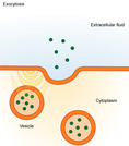
By the end of this section, you will be able to:Describe endocytosis, …
By the end of this section, you will be able to:Describe endocytosis, including phagocytosis, pinocytosis, and receptor-mediated endocytosisUnderstand the process of exocytosis
This resource is a video abstract of a research paper created by …
This resource is a video abstract of a research paper created by Research Square on behalf of its authors. It provides a synopsis that's easy to understand, and can be used to introduce the topics it covers to students, researchers, and the general public. The video's transcript is also provided in full, with a portion provided below for preview:
"White spot syndrome virus (WSSV) is the most destructive virus in crustacean aquaculture, causing huge economic losses. Preventing the initial entry of the virus into host cells is likely the most economical way to control WSSV infection. However, the exact mechanism of virus invasion isn’t clear. To learn more, researchers recently investigated how WSSV evades the host immune system in the crayfish Procambrus clarkii. They found that the enzyme TRIM was significantly upregulated in WSSV-infected crayfish. A recombinant TRIM protein inhibited WSSV replication in the crayfish, while blocking TRIM promoted it, suggesting that this enzyme plays a protective role. Further experiments revealed that TRIM interacts with the viral protein VP26. This interaction keeps the host protein AP-1 from entering the nucleus and driving the expression of dynamin. Without dynamin, WSSV can’t enter the host cell via membrane vesicles..."
The rest of the transcript, along with a link to the research itself, is available on the resource itself.
Phagocytosis is a very primitive system of defence against infection, having even …
Phagocytosis is a very primitive system of defence against infection, having even been shown to exist in invertebrates and single cell organisms. The discovery was made in starfish larvae by Elle Metchnikoff who subsequently won the Nobel Prize for Medicine or Physiology in 1908. The process of phagocytosis itself is a form of endocytosis (cell eating), with vesicular internalisation being the method of removal of pathogens and dead cells (those that have undergone apoptosis, or Programmed Cell Death). This internalised vesicle is referred to as the "phagosome".
This resource is a video abstract of a research paper created by …
This resource is a video abstract of a research paper created by Research Square on behalf of its authors. It provides a synopsis that's easy to understand, and can be used to introduce the topics it covers to students, researchers, and the general public. The video's transcript is also provided in full, with a portion provided below for preview:
"Alzheimer’s disease affects 1 in 3 senior citizens worldwide. The hallmark of Alzheimer’s is the accumulation of abnormal protein deposits, including Tau and amyloid β, in neurons and glial cells in the brain. These deposits disrupt signal transduction by affecting lipid-based secondary messengers in the brain called phosphatidylinositols (PIs). PIs drive the reorganization of the cytoskeleton in glial cells, affecting many cellular processes. These dynamic molecules are tightly regulated by their phosphorylation status, which influences their abundance and localization. Because microglia in the brain must respond to chemotactic and pro-inflammatory signals, disrupting PIs alters microglial function, resulting in hyperactivation and inflammation. PI signaling typically drives actin remodeling to modulate phagocytosis, allowing glial cells to clear amyloid β aggregates and debris. Unfortunately, extensive amyloid β accumulation disrupts PI signaling, altering cytoskeleton regulation..."
The rest of the transcript, along with a link to the research itself, is available on the resource itself.
This resource is a video abstract of a research paper created by …
This resource is a video abstract of a research paper created by Research Square on behalf of its authors. It provides a synopsis that's easy to understand, and can be used to introduce the topics it covers to students, researchers, and the general public. The video's transcript is also provided in full, with a portion provided below for preview:
"New research suggests that cells from the umbilical cord can be programmed to gobble up and kill disease-causing bacteria. When deployed in rats, such cells could effectively reduce signs of acute lung injury, pointing to an alternative route for fighting lung disease in humans. These are mesenchymal stromal cells. Their chameleon-like ability to form into bone, cartilage, or fat in the body has made them valuable for tissue repair and regeneration. But recent studies have shown that these cells can also help boost the immune system. They do this by releasing bioactive pockets of cellular matter that are believed to signal immune cells like macrophages to action. In rats with bacterial lung disease, that ability appears to provide significant relief. Researchers found that injecting mesenchymal cells from human umbilical cords could reduce signs of pneumonia caused by E. coli and increase animal survival. What’s more, they could actually enhance that effect..."
The rest of the transcript, along with a link to the research itself, is available on the resource itself.
This resource is a video abstract of a research paper created by …
This resource is a video abstract of a research paper created by Research Square on behalf of its authors. It provides a synopsis that's easy to understand, and can be used to introduce the topics it covers to students, researchers, and the general public. The video's transcript is also provided in full, with a portion provided below for preview:
"Our bodies must undergo tissue self-renewal in order to remain healthy, and cell death is an important part of self-renewal. Apoptosis is a mechanism of programmed cell death that maintains homeostasis without inflammation. As dying cells begin to dismantle, they signal to phagocytes to engulf them, a process called efferocytosis. The balance between these “find-me,” “eat-me,” and “don’t-eat-me” signals is critical. Unfortunately, because efferocytosis prevents inflammatory responses, these signaling pathways are often hijacked by cancer cells to facilitate immune escape. Although traditional cancer therapies, such as chemotherapy and radiation, kill cancer cells directly, the resulting apoptosis can increase efferocytosis and suppress the immune response, allowing for progression of residual cancer. A new strategy is to combine traditional therapies with those that inhibit efferocytosis, killing tumor cells while blocking the pathways that allow them to proliferate..."
The rest of the transcript, along with a link to the research itself, is available on the resource itself.
This resource is a video abstract of a research paper created by …
This resource is a video abstract of a research paper created by Research Square on behalf of its authors. It provides a synopsis that's easy to understand, and can be used to introduce the topics it covers to students, researchers, and the general public. The video's transcript is also provided in full, with a portion provided below for preview:
"Sepsis is one of the main causes of death in the intensive care unit. When an infection becomes severe, the immune system misfires, triggering a cascade of inflammatory responses. Pro-inflammatory factors secreted by immune cells called macrophages enhance the damage, worsening the clinical picture, and while interventions for sepsis are available, mortality remains high. Now, researchers have identified a new target in the battle against sepsis. Using cells isolated from mice, they measured gene and protein expression during E. coli challenge. They found that cytochrome P450 1A1 (CYP1A1), an enzyme that regulates metabolism, also regulates inflammatory responses during sepsis. CYP1A1 directed macrophages to initiate a microbe-internalizing process, phagocytosis, during infection. Inhibiting CYP1A1 blocked phagocytosis of bacteria in macrophages, preventing the cells from secreting more inflammatory molecules. This strategy was also effective in a mouse model of sepsis..."
The rest of the transcript, along with a link to the research itself, is available on the resource itself.
No restrictions on your remixing, redistributing, or making derivative works. Give credit to the author, as required.
Your remixing, redistributing, or making derivatives works comes with some restrictions, including how it is shared.
Your redistributing comes with some restrictions. Do not remix or make derivative works.
Most restrictive license type. Prohibits most uses, sharing, and any changes.
Copyrighted materials, available under Fair Use and the TEACH Act for US-based educators, or other custom arrangements. Go to the resource provider to see their individual restrictions.