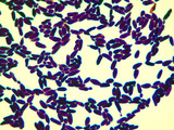
This micrograph was taken at 1000X total magnifcation on a brightfield microscope. …
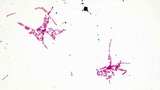
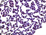
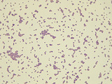
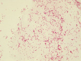
This micrograph was taken at 1000X total magnifcation on a brightfield microscope. The subject is Bacillus cereus cells were grown in broth culture for 48 hours at 30 degrees Celsius. The cells were heat-fixed to a slide and stained with malachite green (endospores) and safranin red (vegetative cells) prior to visualization.Image credit: Emily Fox
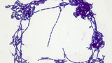
This micrograph was taken at 1000X total magnifcation on a brightfield microscope. …
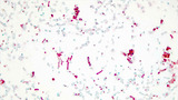
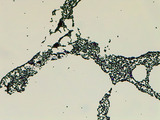
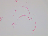
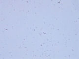
This micrograph was taken at 1000X total magnifcation on a brightfield microscope. The subject is Bacillus megaterium cells were grown in broth culture for 5 days at 30 degrees Celsius. The cells were heat-fixed to a slide and stained with malachite green (endospores) and safranin red (vegetative cells) prior to visualization.Image credit: Emily Fox
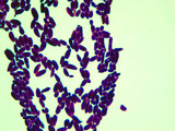
This micrograph was taken at 1000X total magnifcation on a brightfield microscope. …
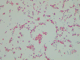
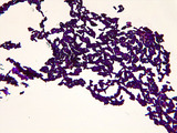
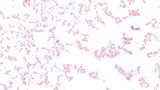
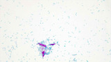
This micrograph was taken at 1000X total magnifcation on a brightfield microscope. The subject is Bacillus subtilis cells were grown in broth culture for 48 hours at 30 degrees Celsius. The cells were heat-fixed to a slide and stained with malachite green (endospores) and safranin red (vegetative cells) prior to visualization.Image credit: Emily Fox
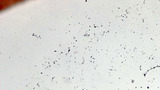
This micrograph was taken at 1000X total magnifcation on a brightfield microscope. …
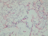
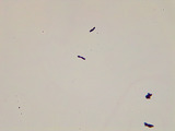
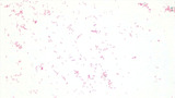
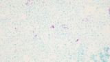
This micrograph was taken at 1000X total magnifcation on a brightfield microscope. The subject is Bacillus subtilis cells were grown in broth culture for 5 days at 30 degrees Celsius. The cells were heat-fixed to a slide and stained with malachite green (endospores) and safranin red (vegetative cells) prior to visualization.Image credit: Emily Fox
This micrograph was taken at 1000X total magnifcation on a brightfield microscope. …
This micrograph was taken at 1000X total magnifcation on a brightfield microscope. The subject is Bacillus subtilis cells grown in broth culture overnight at 30 degrees Celsius. The cells were heat-fixed to a slide Gram stained prior to visualization.Image credit: Emily Fox
This micrograph was taken at 100X total magnifcation on a brightfield microscope. …
This micrograph was taken at 100X total magnifcation on a brightfield microscope. The subject is Bacillus subtilis cells grown in broth culture overnight at 30 degrees Celsius. The cells were heat-fixed to a slide Gram stained prior to visualization.Image credit: Emily Fox
This micrograph was taken at 1000X total magnifcation on a brightfield microscope. …
This micrograph was taken at 1000X total magnifcation on a brightfield microscope. The subject is Candida albicans cells grown in broth culture at 30 degrees Celsius. The cells were heat-fixed to a slide and Gram stained prior to visualization.Image credit: Emily Fox
This micrograph was taken at 1000X total magnifcation on a brightfield microscope. …
This micrograph was taken at 1000X total magnifcation on a brightfield microscope. The subject is Candida albicans cells grown on YPD agar at 30 degrees Celsius. The cells were heat-fixed to a slide and Gram stained prior to visualization.Image credit: Emily Fox
This micrograph was taken at 100X total magnifcation on a brightfield microscope. …
This micrograph was taken at 100X total magnifcation on a brightfield microscope. The subject is Candida albicans cells grown in broth culture at 30 degrees Celsius. The cells were heat-fixed to a slide and Gram stained prior to visualization.Image credit: Emily Fox
This micrograph was taken at 400X total magnifcation on a brightfield microscope. …
This micrograph was taken at 400X total magnifcation on a brightfield microscope. The subject is Candida albicans cells grown in broth culture at 30 degrees Celsius. The cells were heat-fixed to a slide and Gram stained prior to visualization.Image credit: Emily Fox
This micrograph was taken at 1000X total magnifcation on a brightfield microscope. …
This micrograph was taken at 1000X total magnifcation on a brightfield microscope. The subject is unidentified coccus cells from a contaminant colony grown on nutrient agar at 30 degrees Celsius. The cells were heat-fixed to a slide and Gram stained prior to visualization.Image credit: Emily Fox
This micrograph was taken at 1000X total magnifcation on a brightfield microscope. …
This micrograph was taken at 1000X total magnifcation on a brightfield microscope. The subject is Enterobacter aerogenes cells grown in broth culture overnight at 37 degrees Celsius. The cells were heat-fixed to a slide and Gram stained prior to visualization.Image credit: Emily Fox
This micrograph was taken at 1000X total magnifcation on a brightfield microscope. …
This micrograph was taken at 1000X total magnifcation on a brightfield microscope. The subject is Escherichia coli cells were grown in broth culture for 48 hours at 30 degrees Celsius. The cells were heat-fixed to a slide and stained with malachite green (endospores) and safranin red (vegetative cells) prior to visualization. No endospores are seen, as E. coli does not form endospores.Image credit: Emily Fox
This micrograph was taken at 1000X total magnifcation on a brightfield microscope. …
This micrograph was taken at 1000X total magnifcation on a brightfield microscope. The subject is Escherichia coli cells were grown in broth culture for 5 days at 30 degrees Celsius. The cells were heat-fixed to a slide and stained with malachite green (endospores) and safranin red (vegetative cells) prior to visualization. No endospores are seen, as E. coli does not form endospores.Image credit: Emily Fox
This micrograph was taken at 1000X total magnifcation on a brightfield microscope. …
This micrograph was taken at 1000X total magnifcation on a brightfield microscope. The subject is Escherichia coli cells grown in broth culture overnight at 37 degrees Celsius. The cells were heat-fixed to a slide and Gram stained prior to visualization.Image credit: Emily Fox
This micrograph was taken at 400X total magnifcation on a brightfield microscope. …
This micrograph was taken at 400X total magnifcation on a brightfield microscope. The subject is Escherichia coli cells grown in broth culture overnight at 37 degrees Celsius. The cells were heat-fixed to a slide and Gram stained prior to visualization.Image credit: Emily Fox
This micrograph was taken at 1000X total magnifcation on a brightfield microscope. …
This micrograph was taken at 1000X total magnifcation on a brightfield microscope. The subject is Escherichia coli cells and Mycobacterium smegmatis cells grown separately in broth culture at 37 degrees Celsius. The cells were heat-fixed to a slide and acid-fast stained using the Ziehl-Neelsen method prior to visualization. M. smegmatis (acid-fast) stains pink with carbol fuchsin. E.coli (non-acid-fast) stains blue with methylene blue counterstain.Image credit: Emily Fox
No restrictions on your remixing, redistributing, or making derivative works. Give credit to the author, as required.
Your remixing, redistributing, or making derivatives works comes with some restrictions, including how it is shared.
Your redistributing comes with some restrictions. Do not remix or make derivative works.
Most restrictive license type. Prohibits most uses, sharing, and any changes.
Copyrighted materials, available under Fair Use and the TEACH Act for US-based educators, or other custom arrangements. Go to the resource provider to see their individual restrictions.