Biology is designed for multi-semester biology courses for science majors. It is …
Biology is designed for multi-semester biology courses for science majors. It is grounded on an evolutionary basis and includes exciting features that highlight careers in the biological sciences and everyday applications of the concepts at hand. To meet the needs of today’s instructors and students, some content has been strategically condensed while maintaining the overall scope and coverage of traditional texts for this course. Instructors can customize the book, adapting it to the approach that works best in their classroom. Biology also includes an innovative art program that incorporates critical thinking and clicker questions to help students understand—and apply—key concepts.
By the end of this section, you will be able to:Describe the …
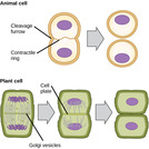
By the end of this section, you will be able to:Describe the three stages of interphaseDiscuss the behavior of chromosomes during karyokinesisExplain how the cytoplasmic content is divided during cytokinesisDefine the quiescent G0 phase
By the end of this section, you will be able to:Describe the …
By the end of this section, you will be able to:Describe the three stages of interphaseDiscuss the behavior of chromosomes during karyokinesisExplain how the cytoplasmic content is divided during cytokinesisDefine the quiescent G0 phase
By the end of this section, you will be able to:Describe the …
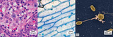
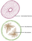
By the end of this section, you will be able to:Describe the cytoskeletonCompare the roles of microfilaments, intermediate filaments, and microtubulesCompare and contrast cilia and flagellaSummarize the differences among the components of prokaryotic cells, animal cells, and plant cells
By the end of this section, you will be able to:List the …
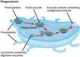
By the end of this section, you will be able to:List the components of the endomembrane systemRecognize the relationship between the endomembrane system and its functions
This resource is a video abstract of a research paper created by …
This resource is a video abstract of a research paper created by Research Square on behalf of its authors. It provides a synopsis that's easy to understand, and can be used to introduce the topics it covers to students, researchers, and the general public. The video's transcript is also provided in full, with a portion provided below for preview:
"FAF1 is a protein involved in various biochemical processes including cell death, inflammation, and cell proliferation and is implicated in certain diseases, including cancer and Parkinson’s disease. To date, FAF1 has been assumed to be locked within the cytosol—with no secretion mechanism reported for the protein. Now, researchers have discovered two mechanisms by which FAF1 can be secreted and transmitted between cells. Experiments on human neuroblastoma cells showed that FAF1 was secreted as cargo within exosomes, as well as in a free, non-exosomal form. Experiments also showed that FAF1 promoted the formation of exosomes, suggesting a regulatory role for the protein in exosome biogenesis. Additionally, extracellular FAF1 was transmitted to neighboring neuronal cells via endocytosis, triggering cell death through apoptotic and necrotic pathways. As the first to reveal these FAF1 secretion pathways this study could lead to ways of interfering with cell death by inhibiting FAF1 secretion..."
The rest of the transcript, along with a link to the research itself, is available on the resource itself.
This resource is a video abstract of a research paper created by …
This resource is a video abstract of a research paper created by Research Square on behalf of its authors. It provides a synopsis that's easy to understand, and can be used to introduce the topics it covers to students, researchers, and the general public. The video's transcript is also provided in full, with a portion provided below for preview:
"The inside of a cell is abuzz with activity… including constant shipments of proteins in membrane-bounded vesicles. Antibodies headed out to the bloodstream to fight disease; enzymes destined for lysosomes to break down and recycle cellular material . But how do all these vesicular parcels get to the right place? It’s already known that long proteins called golgins serve as addresses for and help capture vesicles heading to the Golgi apparatus, the cell’s central sorting station. But little is known about how they do it. Now, researchers at the MRC Laboratory of Molecular Biology in the UK have tracked down the parts of the golgins that act as postal codes. To find out which parts of the golgins provide this critical address function, the team relocated the proteins to the mitochondria, then deleted or mutated different sections to see which sequences were critical for capturing vesicles..."
The rest of the transcript, along with a link to the research itself, is available on the resource itself.
No restrictions on your remixing, redistributing, or making derivative works. Give credit to the author, as required.
Your remixing, redistributing, or making derivatives works comes with some restrictions, including how it is shared.
Your redistributing comes with some restrictions. Do not remix or make derivative works.
Most restrictive license type. Prohibits most uses, sharing, and any changes.
Copyrighted materials, available under Fair Use and the TEACH Act for US-based educators, or other custom arrangements. Go to the resource provider to see their individual restrictions.