This resource is a video abstract of a research paper created by …
This resource is a video abstract of a research paper created by Research Square on behalf of its authors. It provides a synopsis that's easy to understand, and can be used to introduce the topics it covers to students, researchers, and the general public. The video's transcript is also provided in full, with a portion provided below for preview:
"Autophagy is an important cellular recycling process that degrades misfolded proteins and damaged organelles. In typical (“canonical”) autophagy, membranes derived from the endoplasmic reticulum surround damaged materials that need to be degraded, and the proteins Atg5 and Atg7 help form specialized digestion compartments (autophagosomes), but another type of autophagy, called alternative autophagy, was recently discovered. In alternative autophagy, the membranes that envelop the damaged materials are derived from the trans-Golgi membrane, and Atg5 and Atg7 do not participate in autophagosome formation. Alternative autophagy seems to be activated primarily under conditions of cell stress, and it plays roles in many diseases, such as heart disease, neurodegenerative disease, cancer, inflammatory bowel disease, and bacterial infection..."
The rest of the transcript, along with a link to the research itself, is available on the resource itself.
Biology is designed for multi-semester biology courses for science majors. It is …
Biology is designed for multi-semester biology courses for science majors. It is grounded on an evolutionary basis and includes exciting features that highlight careers in the biological sciences and everyday applications of the concepts at hand. To meet the needs of today’s instructors and students, some content has been strategically condensed while maintaining the overall scope and coverage of traditional texts for this course. Instructors can customize the book, adapting it to the approach that works best in their classroom. Biology also includes an innovative art program that incorporates critical thinking and clicker questions to help students understand—and apply—key concepts.
By the end of this section, you will be able to:Describe the …
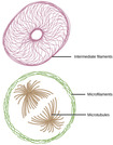
By the end of this section, you will be able to:Describe the cytoskeletonCompare the roles of microfilaments, intermediate filaments, and microtubulesCompare and contrast cilia and flagellaSummarize the differences among the components of prokaryotic cells, animal cells, and plant cells
By the end of this section, you will be able to:List the …
By the end of this section, you will be able to:List the components of the endomembrane systemRecognize the relationship between the endomembrane system and its functions
This resource is a video abstract of a research paper created by …
This resource is a video abstract of a research paper created by Research Square on behalf of its authors. It provides a synopsis that's easy to understand, and can be used to introduce the topics it covers to students, researchers, and the general public. The video's transcript is also provided in full, with a portion provided below for preview:
"Critical defects that compromise the nucleus during cell division could be the basis for the age-accelerating effects of people living with progeria. Hutchinson-Gilford progeria syndrome is a genetic disorder that causes premature aging. Affecting one in 8 million newborns worldwide, the disorder is extremely rare—and fatal. The rapid aging of the cardiovascular system causes death due to heart attack or stroke in patients by their mid-teens. Progeria is caused by a tiny point mutation in the lamin A gene. This gene is responsible for producing structural proteins called lamins, which form the scaffolding that holds the cell nucleus together. The mutated form of prelamin A called progerin destabilizes the cell nucleus—the genetic control center of cells. The result is the fast-aging effects observed in progeria. But the link from gene mutation to physical disorder has remained a mystery. Previous studies have looked only at models of progeria, not at actual patient cells..."
The rest of the transcript, along with a link to the research itself, is available on the resource itself.
This resource is a video abstract of a research paper created by …
This resource is a video abstract of a research paper created by Research Square on behalf of its authors. It provides a synopsis that's easy to understand, and can be used to introduce the topics it covers to students, researchers, and the general public. The video's transcript is also provided in full, with a portion provided below for preview:
"Alzheimer’s disease is a progressive brain disorder that gradually destroys memory and thinking skills Every year, the number of people affected by the disease continues to grow That has some researchers looking to the fruit fly for answers One team has found that linking two parts of the cell closer together may help Linking the endoplasmic reticulum, which forms proteins and stores calcium to the mitochondria, which power the cell can actually improve motor function in fruit flies and help them live longer This technique works in flies with brain plaques similar to those found in humans with Alzheimer’s disease Part of the reason could be improved access to calcium Forcing the organelles together helps calcium migrate more easily from the endoplasmic reticulum to the mitochondria This sends the mitochondria into overdrive because calcium acts as a lubricant for the mitochondrial machinery that pumps out energy So easy access to calcium means more energy output Clarifying how that transl.."
The rest of the transcript, along with a link to the research itself, is available on the resource itself.
This resource is a video abstract of a research paper created by …
This resource is a video abstract of a research paper created by Research Square on behalf of its authors. It provides a synopsis that's easy to understand, and can be used to introduce the topics it covers to students, researchers, and the general public. The video's transcript is also provided in full, with a portion provided below for preview:
"Calcium ions (Ca²⁺) are critical secondary messengers in our cells, shuttling messages from outside cells to within. Ca²⁺ signaling depends on transport proteins to move ions across membranes. Store-operated channels (SOCs) are one particularly important class of transporters for Ca²⁺ signaling. SOCs allow Ca²⁺ signaling to continue by refilling critical Ca²⁺ stores in a process called store-operated Ca²⁺ entry (SOCE). Our cardiovascular system is particularly dependent on Ca²⁺ signaling, even in non-excitable (i.g., non-muscle) cells like vascular endothelial cells. Growing evidence suggests that malfunctions in SOCs and the SOCE process contribute to many cardiovascular diseases. But the exact roles of SOCs and SOCE are not fully understood. A recent review examined the current literature on SOC function in the vasculature and what is currently known about SOCs' role in cardiovascular disease..."
The rest of the transcript, along with a link to the research itself, is available on the resource itself.
This resource is a video abstract of a research paper created by …
This resource is a video abstract of a research paper created by Research Square on behalf of its authors. It provides a synopsis that's easy to understand, and can be used to introduce the topics it covers to students, researchers, and the general public. The video's transcript is also provided in full, with a portion provided below for preview:
"FAF1 is a protein involved in various biochemical processes including cell death, inflammation, and cell proliferation and is implicated in certain diseases, including cancer and Parkinson’s disease. To date, FAF1 has been assumed to be locked within the cytosol—with no secretion mechanism reported for the protein. Now, researchers have discovered two mechanisms by which FAF1 can be secreted and transmitted between cells. Experiments on human neuroblastoma cells showed that FAF1 was secreted as cargo within exosomes, as well as in a free, non-exosomal form. Experiments also showed that FAF1 promoted the formation of exosomes, suggesting a regulatory role for the protein in exosome biogenesis. Additionally, extracellular FAF1 was transmitted to neighboring neuronal cells via endocytosis, triggering cell death through apoptotic and necrotic pathways. As the first to reveal these FAF1 secretion pathways this study could lead to ways of interfering with cell death by inhibiting FAF1 secretion..."
The rest of the transcript, along with a link to the research itself, is available on the resource itself.
The MIT Biology Department core courses, 7.012, 7.013, and 7.014, all cover …
The MIT Biology Department core courses, 7.012, 7.013, and 7.014, all cover the same core material, which includes the fundamental principles of biochemistry, genetics, molecular biology, and cell biology. Biological function at the molecular level is particularly emphasized and covers the structure and regulation of genes, as well as, the structure and synthesis of proteins, how these molecules are integrated into cells, and how these cells are integrated into multicellular systems and organisms. In addition, each version of the subject has its own distinctive material. 7.012 focuses on the exploration of current research in cell biology, immunology, neurobiology, genomics, and molecular medicine. Acknowledgments The study materials, problem sets, and quiz materials used during Fall 2004 for 7.012 include contributions from past instructors, teaching assistants, and other members of the MIT Biology Department affiliated with course #7.012. Since the following works have evolved over a period of many years, no single source can be attributed.
The MIT Biology Department core Introductory Biology courses, 7.012, 7.013, 7.014, 7.015, …
The MIT Biology Department core Introductory Biology courses, 7.012, 7.013, 7.014, 7.015, and 7.016 all cover the same core material, which includes the fundamental principles of biochemistry, genetics, molecular biology, and cell biology. The focus of 7.013 is on genomic approaches to human biology, including neuroscience, development, immunology, tissue repair and stem cells, tissue engineering, and infectious and inherited diseases, including cancer.
The MIT Biology Department core courses, 7.012, 7.013, and 7.014, all cover …
The MIT Biology Department core courses, 7.012, 7.013, and 7.014, all cover the same core material, which includes the fundamental principles of biochemistry, genetics, molecular biology, and cell biology. Biological function at the molecular level is particularly emphasized and covers the structure and regulation of genes, as well as, the structure and synthesis of proteins, how these molecules are integrated into cells, and how these cells are integrated into multicellular systems and organisms. In addition, each version of the subject has its own distinctive material. 7.014 focuses on the application of these fundamental principles, toward an understanding of microorganisms as geochemical agents responsible for the evolution and renewal of the biosphere and of their role in human health and disease. Acknowledgements The study materials, problem sets, and quiz materials used during Spring 2005 for 7.014 include contributions from past instructors, teaching assistants, and other members of the MIT Biology Department affiliated with course 7.014. Since the following works have evolved over a period of many years, no single source can be attributed.
The MIT Biology Department core courses, 7.012, 7.013, and 7.014, all cover …
The MIT Biology Department core courses, 7.012, 7.013, and 7.014, all cover the same core material, which includes the fundamental principles of biochemistry, genetics, molecular biology, and cell biology. 7.013 focuses on the application of the fundamental principles toward an understanding of human biology. Topics include genetics, cell biology, molecular biology, disease (infectious agents, inherited diseases and cancer), developmental biology, neurobiology and evolution. Biological function at the molecular level is particularly emphasized in all courses and covers the structure and regulation of genes, as well as, the structure and synthesis of proteins, how these molecules are integrated into cells, and how these cells are integrated into multicellular systems and organisms. In addition, each version of the subject has its own distinctive material.
This resource is a video abstract of a research paper created by …
This resource is a video abstract of a research paper created by Research Square on behalf of its authors. It provides a synopsis that's easy to understand, and can be used to introduce the topics it covers to students, researchers, and the general public. The video's transcript is also provided in full, with a portion provided below for preview:
"Multiple myeloma (MM) is a common type of plasma cell cancer that remains aggressive and incurable despite the development of several treatments. Approximately 70–80% of patients with MM have myeloma bone disease, which involves bone fractures and high blood calcium (Ca2+) and affects MM prognosis. Various calcium channels and transporters help balance calcium levels, so they may be closely related to MM prognosis. For example, plasma membrane calcium channels allow calcium ions to enter cells, while proteins involved in store-operated calcium entry (SOCE) mediate calcium release from sites in the endoplasmic reticulum (ER)/sarcoplasmic reticulum (SR). Mitochondrial calcium channels regulate calcium uptake into mitochondria, which contributes to SOCE, and calcium-ATPases pump calcium ions from the cytoplasm back into the ER/SR or extracellular space. These molecules have been reported to be altered in the context of MM, but the specific mechanism by which their dysfunction leads to MM remains unclear..."
The rest of the transcript, along with a link to the research itself, is available on the resource itself.
This resource is a video abstract of a research paper created by …
This resource is a video abstract of a research paper created by Research Square on behalf of its authors. It provides a synopsis that's easy to understand, and can be used to introduce the topics it covers to students, researchers, and the general public. The video's transcript is also provided in full, with a portion provided below for preview:
"Autophagy is the process by which healthy cells degrade and recycle waste material. Researchers are finding that this vital function is interrupted in different forms of cancer, including brain cancer. A new review describes how researchers are repairing broken autophagy pathways in tumors using microRNAs, or miRNAs. miRNAs are small non-coding RNA molecules that regulate a variety of cellular processes— including autophagy. Understanding the molecular targets of miRNAs and their function is crucial, as it could lead to the development of new therapies for patients with brain tumors..."
The rest of the transcript, along with a link to the research itself, is available on the resource itself.
This resource is a video abstract of a research paper created by …
This resource is a video abstract of a research paper created by Research Square on behalf of its authors. It provides a synopsis that's easy to understand, and can be used to introduce the topics it covers to students, researchers, and the general public. The video's transcript is also provided in full, with a portion provided below for preview:
"The development of solid tumors like melanomas is driven by cancer stem cells (CSCs). These cells can also promote tumor growth, dormancy, metastasis, recurrence, and chemoresistance, which contribute to poor cancer outcomes. An intracellular degradation/recycling pathway called autophagy is thought to regulate the “stemness” of CSCs to enable these effects. To clarify the mechanism, a recent study examined CSCs isolated from human melanoma cell lines. The expression of the molecule Sec23a was lower in the CSCs than in the original melanoma cells, and the reduced Sec23a expression was associated with increased stemness in vitro and tumor growth in vivo, indicating a negative correlation between Sec23a and CSC stemness. Further experiments revealed that Sec23a downregulation increases CSC stemness by promoting autophagy. Specifically, Sec23a downregulation enhances endoplasmic reticulum (ER) stress, which leads to upregulation of the ER stress-responsive protein FAM134B..."
The rest of the transcript, along with a link to the research itself, is available on the resource itself.
This resource is a video abstract of a research paper created by …
This resource is a video abstract of a research paper created by Research Square on behalf of its authors. It provides a synopsis that's easy to understand, and can be used to introduce the topics it covers to students, researchers, and the general public. The video's transcript is also provided in full, with a portion provided below for preview:
"The endoplasmic reticulum (ER) ensures that newly synthesized proteins in a cell are properly folded. But when the number of new proteins exceeds the ER’s capacity to fold them, ER stress can occur and the “unfolded protein response” (UPR) is triggered to help return the cell back to normal. If the UPR can’t restore this balance, the cell dies. Triggering cell death by ER stress via the UPR is one way to treat diseases such as cancer, but we must fully understand the signaling mechanisms involved to design drugs to effectively trigger the UPR. ER stressors such as thapsigargin can induce UPR-mediated cell death, but the detailed mechanisms are unclear. In a recent study, researchers sought to better understand how ER stressors work. They analyzed molecular changes in human prostate and colon cancer cell lines exposed to thapsigargin or its analogs. Their results demonstrated which UPR components and cell death processes are required for an ER stressor to effectively kill a cell..."
The rest of the transcript, along with a link to the research itself, is available on the resource itself.
This resource is a video abstract of a research paper created by …
This resource is a video abstract of a research paper created by Research Square on behalf of its authors. It provides a synopsis that's easy to understand, and can be used to introduce the topics it covers to students, researchers, and the general public. The video's transcript is also provided in full, with a portion provided below for preview:
"Chronic infection with the hepatitis C virus is a major cause of chronic liver disease, even after the virus has been eradicated by antiviral treatment. The problem appears to lie in the lingering activation of harmful Wnt/β-catenin signaling, which active viruses exploit for replication. A new study suggests the enzyme PKA could play a role. PKA is part of a signaling cascade that is activated during hepatitis C infection. To determine its role, researchers prevented PKA activation by treating cells with a PKA inhibitor. Inhibition was found to be beneficial. Inhibiting PKA reduced cells’ capacity to support both the hepatitis C virus and Wnt/β-catenin signaling, as mediated by another enzyme, GSK-3β. Interestingly, similar benefits were observed when another harmful effect of viral infection was repressed, namely, endoplasmic reticulum stress..."
The rest of the transcript, along with a link to the research itself, is available on the resource itself.
No restrictions on your remixing, redistributing, or making derivative works. Give credit to the author, as required.
Your remixing, redistributing, or making derivatives works comes with some restrictions, including how it is shared.
Your redistributing comes with some restrictions. Do not remix or make derivative works.
Most restrictive license type. Prohibits most uses, sharing, and any changes.
Copyrighted materials, available under Fair Use and the TEACH Act for US-based educators, or other custom arrangements. Go to the resource provider to see their individual restrictions.