This resource is a video abstract of a research paper created by …
This resource is a video abstract of a research paper created by Research Square on behalf of its authors. It provides a synopsis that's easy to understand, and can be used to introduce the topics it covers to students, researchers, and the general public. The video's transcript is also provided in full, with a portion provided below for preview:
"Delirium, or a confused or disoriented state that can affect attention, awareness and cognition, is common in elderly patients following surgery -- and is a leading cause of postoperative complications among elderly hospitalized patients. But it’s not clear why this happens or which patients are at especially high risk. In particular, certain electroencephalogram (E-E-G) patterns during anesthesia known as burst-suppression have been associated with postoperative delirium. These patterns are characterized by spikes in electrical activity, or bursts, alternating with longer periods of no activity. But whether burst-suppression plays a causal role in delirium isn’t known. A study, now published in the journal Anesthesiology by researchers in Boston, finds that in elderly patients undergoing cardiopulmonary bypass, burst-suppression is associated with delirium. The project was a sub-study of the ongoing MINDDS trial, and retrospectively looked at the outcomes of 159 patients over the age of 60..."
The rest of the transcript, along with a link to the research itself, is available on the resource itself.
This resource is a video abstract of a research paper created by …
This resource is a video abstract of a research paper created by Research Square on behalf of its authors. It provides a synopsis that's easy to understand, and can be used to introduce the topics it covers to students, researchers, and the general public. The video's transcript is also provided in full, with a portion provided below for preview:
"Scientists have uncovered new information on how sensory processing can change in multiple sclerosis, or MS. Their findings could open novel avenues for understanding and treating the disease. People with MS typically experience deficits in their ability to smell and taste, abnormal temperature processing, and heightened sensations of pain or fatigue. But there’s no clear neurocognitive mechanism to explain such diverse symptoms. A new report in the journal Human Brain Mapping suggests these changes may stem from a problem with interoception. Interoception is a lesser-known skill that helps people feel signals originating from inside of the body – such as the heart beating or the digestive system signaling hunger. Many of the sensory processing issues that characterize MS are associated with brain pathways related to interoception. This link prompted researchers to examine how these pathways are affected in the context of the disease..."
The rest of the transcript, along with a link to the research itself, is available on the resource itself.
Psychology is designed to meet scope and sequence requirements for the single-semester …
Psychology is designed to meet scope and sequence requirements for the single-semester introduction to psychology course. The book offers a comprehensive treatment of core concepts, grounded in both classic studies and current and emerging research. The text also includes coverage of the DSM-5 in examinations of psychological disorders. Psychology incorporates discussions that reflect the diversity within the discipline, as well as the diversity of cultures and communities across the globe.Senior Contributing AuthorsRose M. Spielman, Formerly of Quinnipiac UniversityContributing AuthorsKathryn Dumper, Bainbridge State CollegeWilliam Jenkins, Mercer UniversityArlene Lacombe, Saint Joseph's UniversityMarilyn Lovett, Livingstone CollegeMarion Perlmutter, University of Michigan
By the end of this section, you will be able to:Explain the …
By the end of this section, you will be able to:Explain the functions of the spinal cordIdentify the hemispheres and lobes of the brainDescribe the types of techniques available to clinicians and researchers to image or scan the brain
This resource is a video abstract of a research paper created by …
This resource is a video abstract of a research paper created by Research Square on behalf of its authors. It provides a synopsis that's easy to understand, and can be used to introduce the topics it covers to students, researchers, and the general public. The video's transcript is also provided in full, with a portion provided below for preview:
"How humans lose consciousness under anesthesia is a bit of a mystery, but that’s especially true in the case of young infants. Scientists have known that in adults, fast alpha-wave frequencies in the electroencephalogram, or EEG, appear when someone undergoes anesthesia and loses consciousness. But that doesn’t happen in infants younger than about 4 months. So, how do young infants lose consciousness? Because awake infants do show slow delta-wave frequencies, a team at the University of Cambridge and Harvard Medical School and Boston Children’s Hospital decided to investigate those waves. Now, in a new article in the journal Anesthesiology, the researchers report that infants lost slow-wave synchronization—or “functional connectivity”—between the frontal and parietal regions of their brains when they underwent anesthesia. There is evidence that during anesthesia, the adult brain reorganizes itself into a less complex configuration with more segregation between functional systems..."
The rest of the transcript, along with a link to the research itself, is available on the resource itself.
This resource is a video abstract of a research paper created by …
This resource is a video abstract of a research paper created by Research Square on behalf of its authors. It provides a synopsis that's easy to understand, and can be used to introduce the topics it covers to students, researchers, and the general public. The video's transcript is also provided in full, with a portion provided below for preview:
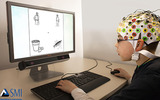
"Understanding how human decision-making and preferences manifest before conscious thought has long challenged researchers focused on cognitive and information science. Now, the field of neuromarketing – a discipline that looks at the neurocognitive underpinnings of consumer behavior – is starting to uncover, in amazing detail, exactly how the brain goes about recognizing a brand. An international research team based in Auckland University of Technology and Nottingham Trent University has devised a new machine learning method that tracks brain responses to logos on the millisecond timescale…even before conscious thoughts are formed. Their results shed light on the early spikes in brain activity that are tied to brand awareness. The method utilizes one of the most promising recent trends in artificial intelligence research: spiking neural networks. These networks use algorithms loosely modeled on the behavior of the human brain to recognize patterns in sets of streaming data..."
The rest of the transcript, along with a link to the research itself, is available on the resource itself.
This resource is a video abstract of a research paper created by …
This resource is a video abstract of a research paper created by Research Square on behalf of its authors. It provides a synopsis that's easy to understand, and can be used to introduce the topics it covers to students, researchers, and the general public. The video's transcript is also provided in full, with a portion provided below for preview:
"Spinal cord stimulation offers long-term relief to patients managing chronic pain, without the need for opioids. This winning combination has spurred incredible growth in the industry, with approximately 50,000 spinal cord stimulators being implanted each year and an estimated market of 7 billion U.S. dollars by 2020. But despite this progress, exactly how the technology works remains unclear. Especially with regard to potential changes in brain activity. A new review article in the journal Anesthesiology takes a closer look at these potential supraspinal pathways and actions. Covering both preclinical and clinical articles, the review starts with the 1960s, when an idea known as Gate Control Theory was proposed to explain how conventional tonic stimulation works. The theory suggests that a combination of presynaptic inhibition and inhibitory interneuronal communication occurs in the spinal cord following the electrical activation of large-diameter afferent fibers..."
The rest of the transcript, along with a link to the research itself, is available on the resource itself.
Systems Neuroscience Laboratory consists of a series of laboratories designed to give …
Systems Neuroscience Laboratory consists of a series of laboratories designed to give students experience with basic techniques for conducting systems neuroscience research. It includes sessions on anatomical, neurophysiological, and data acquisition and analysis techniques, and the ways these techniques are used to study nervous system function. Training is provided in the art of scientific writing with feedback designed to improve writing skills. Assignments include weekly preparation for lab sessions, two major research reports and a series of basic computer programming tutorials (MATLAB). The class involves the use of experimental animals. Enrollment is limited.
No restrictions on your remixing, redistributing, or making derivative works. Give credit to the author, as required.
Your remixing, redistributing, or making derivatives works comes with some restrictions, including how it is shared.
Your redistributing comes with some restrictions. Do not remix or make derivative works.
Most restrictive license type. Prohibits most uses, sharing, and any changes.
Copyrighted materials, available under Fair Use and the TEACH Act for US-based educators, or other custom arrangements. Go to the resource provider to see their individual restrictions.