Deglutition is the process of swallowing. Food is passed from the oral …
Deglutition is the process of swallowing. Food is passed from the oral pharynx into the oesophageal/laryngeal pharynx whilst the epiglottis closes across the entrance of the trachea. It is an involuntary reflex preventing food from passing into the trachea and thus preventing choking and respiratory pneumonia.
Embryo, when applied to mammals, is the term given to the developing …
Embryo, when applied to mammals, is the term given to the developing organism from fertilisation to birth. Developmental biology, or embryology, is the study of the embryo as it transforms from a unicellular zygote to a multicellular, mulitsystemed organism which in some cases is ready to function autonomously at birth. Developmental biology is of interest to vets in understanding why organs and systems are the way they are, but also in understanding genetic diseases and applying cell based therapies to treat loss or damage to tissues.
The Diaphragm is a dome-shaped musculotendinous sheet separating the thoracic and abdominal …
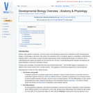
The Diaphragm is a dome-shaped musculotendinous sheet separating the thoracic and abdominal cavities. It is convex on its cranial surface. In the neutral position between full inspiration and full expiration, the most cranial part of the diaphragm is in line with the 6th rib.
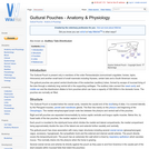
Gases or liquids can be unevenly distributed between two areas. If one …
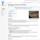
Gases or liquids can be unevenly distributed between two areas. If one area has a higher concentration than the other then the differance between these two areas is termed the concentration gradient. The equality is then corrected by the movement of the molecules down this so called gradient from the region of high concentration to that of low. This process is passive as the molecules do not have to be forced to do this and it is reffered to as diffusion.
The duodenum is the proximal part of the small intestine and extends …
The duodenum is the proximal part of the small intestine and extends from the pylorus of the stomach to the jejunum. It has descending and ascending portions and both portions have digestive and absorptive functions.
Elephant anatomy is very much comparable to the horse and rabbit. Microbes …
Elephant anatomy is very much comparable to the horse and rabbit. Microbes are present in the hindgut that produce Volatile Fatty Acids (VFAs). VFAs make a substantial contribution to the elephant's total energy requirements. Food has a relatively fast transit time and as a result, elephants have a low digestive efficiency (44% as opposed to 60% in horses). A fast transit time is achieved by a short GIT, reduced caecum and increased GIT diameter. Their digestive strategy is to pass as large a quantity of low quality food through their digestive tract within a short period of time.
There are various hormones that influence the structure of the skin. These …
There are various hormones that influence the structure of the skin. These influences may be made apparent by the repeated long-term administration of various glucocorticoids or their analogues. Endogenous imbalances are generally seen in adult mature animals although congenital forms have been seen, especially with hypothyroidism. The hormones implicated as important for maintaining skin structure are thyroxine, cortisol and estradiol. Deficiencies or excessive production may result from abberations in the function of the hypothalamic-adrenal axis, the adrenal gland, thyroid gland or the gonads.
Comprised of a group of duct-less glands with limited or no anatomical …
Comprised of a group of duct-less glands with limited or no anatomical contact with each other, the endocrine system integrates and controls metabolic activity through the secretion of hormones into the vascular system. These hormones may have their effects on tissues and organs far from where they were produced.
Prior to birth the foetus is not capable of respiratory function and …
Prior to birth the foetus is not capable of respiratory function and thus relies on the maternal circulation to carry out gas, nutrient and waste exchange. The foetal and maternal blood never mix, instead they interface at the placenta. Consequently the liver and the lungs are non-functional, and a series of shunts exist in the foetal circulation so that these organs are almost completely by-passed.
The forebrain (proencephalon) is the largest part of the brain, most of …
The forebrain (proencephalon) is the largest part of the brain, most of which is cerebrum. Other important structures found in the forebrain include the thalamus , the hypothalamus and the limbic system. The cerebrum is divided into two cerebral hemispheres connected by a mass of white matter known as the corpus callosum.
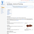
The gall bladder stores bile produced in the liver. Bile is important …
The gall bladder stores bile produced in the liver. Bile is important in the digestion of lipids. The gall bladder forms as an outgrowth of the bile duct, as a secondary hollow at the posterior edge of the original hepatic rudiment. The cystic duct joins the common bile duct which enters the duodenum at the major duodenal papillae (with the pancreatic duct) on the dorsal surface of the duodenum.
The air in the alveoli is renewed regularly, thanks to the ventilation …
The air in the alveoli is renewed regularly, thanks to the ventilation process. Gas exchange in the lungs takes place between the blood in the capillary network surrounding the alveoli, and the air in the alveoli itself.
Gastrulation is the process of forming the three germ layers; ectoderm, mesoderm …
Gastrulation is the process of forming the three germ layers; ectoderm, mesoderm and endoderm. It is achieved through a series of highly coordinated cell movements. Cells that will form the endodermal and mesodermal organs are brought inside the embryo, whilst cells that will form ectoderm move to spread out over the outside of the embryo.
The endoderm will form the lining of the gut and the organs …
The endoderm will form the lining of the gut and the organs that develop from it. Splanchnic mesoderm surrounds the endoderm and orginates from the lateral plate mesoderm. It will form the smooth muscle of the gut that are used in peristalsis.
The Guttural Pouch is present only in members of the order Perissodactyla …
The Guttural Pouch is present only in members of the order Perissodactyla (nonruminant ungulates: horses, tapirs, rhinoceros) and another small band of small mammals including Hyraxes, certain bats and a South American mouse.
Hair germs begin from an aggregation of keratinocytes in the stratum basale …
Hair germs begin from an aggregation of keratinocytes in the stratum basale of the epidermis. The initiating factor is the underlying dermal fibroblast cells. The keratinocytes elongate, divide and relocate to the dermis. Dermal fibroblasts then form a dermal papilla beneath the hair germ. This causes stimulation of the basal stem cells to up-regulate their cycle, producing cells that will keratinise and form the hair shaft. Two swellings form on the shaft, one containing stem cells for follicle regeneration, the other becomes a sebaceous gland which will secrete sebum onto the hair shaft. The follicles develop from an ectodermal bud which invades the mesenchyme during embryonic development. The mesoderm also condenses during the development creating an outer mesodermal component to the embedded part of the hair.
Hair germs begin from an aggregation of keratinocytes in the stratum basale …
Hair germs begin from an aggregation of keratinocytes in the stratum basale of the epidermis. The initiating factor is the underlying dermal fibroblast cells. The keratinocytes elongate, divide and relocate to the dermis. Dermal fibroblasts then form a dermal papilla beneath the hair germ. This causes stimulation of the basal stem cells to up-regulate their cycle, producing cells that will keratinise and form the hair shaft.
The formation of the mammalian heart is a fairly complex process. It …
The formation of the mammalian heart is a fairly complex process. It begins when angiogenic mesodermal cells in the cardiogenic plate coalesce to form the endocardial tubes. The endocardial tubes then fuse to form a single duct, the cardiac tube. This undergoes a process of distension, folding and septation and a four chambered, dual circuit pump is formed . The simple heart seen in fish or amphibians forms via the same path but development ceases at an earlier stage.
No restrictions on your remixing, redistributing, or making derivative works. Give credit to the author, as required.
Your remixing, redistributing, or making derivatives works comes with some restrictions, including how it is shared.
Your redistributing comes with some restrictions. Do not remix or make derivative works.
Most restrictive license type. Prohibits most uses, sharing, and any changes.
Copyrighted materials, available under Fair Use and the TEACH Act for US-based educators, or other custom arrangements. Go to the resource provider to see their individual restrictions.