Also referred to as the reticuloendothelial system or mononuclear phagocytic system. It …
Also referred to as the reticuloendothelial system or mononuclear phagocytic system. It is comprised of primary lymphoid organs (bone marrow, Bursa of Fabricius, the foetal liver and the thymus) which are responsible for the production of lymphocytes, and the secondary lymphoid organs (lymph nodes, spleen and mucosal associated lymphoid tissue) which function to provide an environment where lymphocytes can react to antigen from the tissue fluid, blood and mucosal surfaces.
The musculoskeletal system includes bones, joints, cartilage, muscles, ligaments and tendons. In …
The musculoskeletal system includes bones, joints, cartilage, muscles, ligaments and tendons. In order to describe anatomical landmarks for example for the purposes of surgery and to be able to describe different directional information, for example when recording the view of a recently taken x-ray, it is necessary to have a way of describing the planes and axes that can be applied to the musculoskeletal system to pinpoint a specific anatomical area.
This page has links to information about pregnancy and parturation; including sperm …
This page has links to information about pregnancy and parturation; including sperm in the female tract, fertilisation, sexual differentiation, genital development, gestation lengths in different species, maternal recognition of pregnancy, litter sizes, placenta and its endocrine function, fetal circulation, puerperium, and reproductive disorders.
The Central Nervous System (CNS) is composed of the brain and the …
The Central Nervous System (CNS) is composed of the brain and the spinal cord. This page is specifically focussed on the histologic appearance, for anatomy see Forebrain, Midbrain, Hindbrain, Cranial Nerves, Spinal Cord and CNS Development.
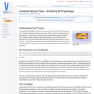
Cerebrospinal fluid (CSF) surrounds the brain as well as the central canal …
Cerebrospinal fluid (CSF) surrounds the brain as well as the central canal of the spinal cord. It helps cushion the central nervous system (CNS), acting in a similar manner to a shock absorber. It also acts as a chemical buffer providing immunological protection and a transport system for waste products and nutrients. The CSF also provides buoyancy to the soft neural tissues which effectively allows the neural tissue to "float" in the CSF. This prevents the brain tissue from becoming deformed under its own weight. It acts as a diffusion medium for the transport of neurotransmitters and neuroendocrine substances.
The cervix can be palpated transrectally and forms a sphincter controlling access …
The cervix can be palpated transrectally and forms a sphincter controlling access to the uterus.The anatomy of the cervical canal is adapted to suit a particular pattern of reproduction and its composition will alter under the influence of reproductive hormones. Not only does it respond to the fluctuation in oestrodiol during the oestrous cycle, but is responsive to prostaglandins and oxytocin in order to 'soften' for parturition.
It is important that all aspects of haemostasis can be independently evaluated. …
It is important that all aspects of haemostasis can be independently evaluated. This will help to identify the phase affected and to pinpoint what the abnormality is. There are tests available to assess primary haemostasis, secondary haemostasis and fibrinolysis.
The colon is a site of microbial fermentation, the relative importance of …
The colon is a site of microbial fermentation, the relative importance of this is species dependent. The colon can be divided into the following portions; Ascending, transverse and descending.
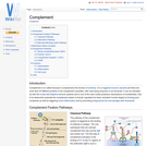
Complement is so called because it complements the function of antibody. It …
Complement is so called because it complements the function of antibody. It is a triggered enzyme cascade and there are more than 20 different proteins in the complement cascades, with most being enzymes or pro-enzymes. It can be activated by both the innate and adaptive immune systems and is one of the main innate protective mechanisms of invertebrates. Due to its destructive potential the complement system is heavily regulated but when activated it works largely by forming pore complexes as well as triggering acute inflammation and by promoting phagocytosis by macrophages and neutrophils.
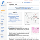
The issue of confounding is of central importance in any analytic epidemiological …
The issue of confounding is of central importance in any analytic epidemiological study (as well as in those descriptive studies aiming to compare different populations), especially in the case of observational studies. Confounding results from non-random differences between the groups of animals being compared in relation to a second, 'confounding' exposure which is independently associated with both the exposure of interest (although not a consequence of this) and the outcome of interest (although not an effect of this). This results in the effect of the exposure of interest is 'mixed up' with the effect of the confounding exposure, and therefore an incorrect estimate of the true association. As such, confounding is viewed by many authors as a form of bias - however, unlike forms of selection and information bias, it is a natural feature of the data (in the case of an observational study), and techniques are available to account for it during analysis.
Different hormones, neurotransmitters and reflexes are involved in the complicated process of …
Different hormones, neurotransmitters and reflexes are involved in the complicated process of feeding in animals. Secretions and motility of the gastrointestinal tract are stimulated and carefully regulated by numerous factors, including environmental stimuli and the presence of food in different parts of the gastrointestinal tract from the oral cavity right through to the intestines. When a harmful substance is ingested the body acts to eliminate it in different ways to prevent the animal becoming ill, for example, through vomiting and diarrhoea. If one or more of the pathways in controlling feeding is damaged or inhibited, then problems such as obesity occurs.
Corpus Luteum is latin for "yellow body". The corpus luteum is the …
Corpus Luteum is latin for "yellow body". The corpus luteum is the structure formed during luteinisation of the follicle after ovulation. The corpus luteum is, however, actually only yellow in the cow and in all other domestic species it is red. The yellow colouration of the corpus luteum is due to the pigment, lutein.
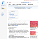
Luteinisation occurs after ovulation and the collapse of the follicle. The number …
Luteinisation occurs after ovulation and the collapse of the follicle. The number of corpora lutea formed in the ovary at any one time is directly proportional to the number of oocytes ovulated. Therefore many corpora lutea will be visible on the ovary of polytocous animals. During Luteinisation there is an increase in both the size and weight due to hyperplasia (increase in cell number) and hypertrophy (increase in cell size) within the developing corpus luteum.
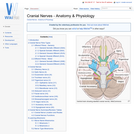
Cranial nerves are those nerves which arise from the brain and brain …
Cranial nerves are those nerves which arise from the brain and brain stem rather than the spinal cord. Nerves arising from the spinal cord are the peripheral nerves. There are 12 pairs of cranial nerves and these pairs of nerves passage through foramina in the skull, either individually or in groups. Cranial nerves are traditionally referred to by Roman numerals and these numerals begin cranially and run caudally.
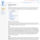
All epidemiological investigations require some form of data description. A number of …
All epidemiological investigations require some form of data description. A number of methods are available for describing data, and the most appropriate one will depend upon both the type of data available and the aims of the investigation. If these issues are not considered, useful information may be lost, or more seriously, a misleading estimate may be made.
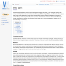
Epidemiological investigation requires a good understanding of different data types, as this …
Epidemiological investigation requires a good understanding of different data types, as this will strongly influence data analysis and interpretation. Data can broadly be classified as qualitative and quantitative, and within each of these groups, data can be further categorised as shown below. Although different grouping systems are available, it is important to consider the type of data being dealt with prior to any analysis. If desired, data can often be changed into different types through manipulation (for example, the quantitative variable weight can be converted to qualitative variables such as low/medium/high or low/not low).
Deglutition is the process of swallowing. Food is passed from the oral …
Deglutition is the process of swallowing. Food is passed from the oral pharynx into the oesophageal/laryngeal pharynx whilst the epiglottis closes across the entrance of the trachea. It is an involuntary reflex preventing food from passing into the trachea and thus preventing choking and respiratory pneumonia.
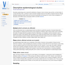
Descriptive epidemiology aims to describe the distribution of disease in terms of …
Descriptive epidemiology aims to describe the distribution of disease in terms of animal, place and time, as shown below. In a purely descriptive study, no attempt is made to formally investigate reasons for the patterns of disease observed, although hypotheses regarding possible reasons will commonly be generated and developed as a result of these investigations. A description of the different types of descriptive studies is provided elsewhere.
Embryo, when applied to mammals, is the term given to the developing …
Embryo, when applied to mammals, is the term given to the developing organism from fertilisation to birth. Developmental biology, or embryology, is the study of the embryo as it transforms from a unicellular zygote to a multicellular, mulitsystemed organism which in some cases is ready to function autonomously at birth. Developmental biology is of interest to vets in understanding why organs and systems are the way they are, but also in understanding genetic diseases and applying cell based therapies to treat loss or damage to tissues.
The Diaphragm is a dome-shaped musculotendinous sheet separating the thoracic and abdominal …
The Diaphragm is a dome-shaped musculotendinous sheet separating the thoracic and abdominal cavities. It is convex on its cranial surface. In the neutral position between full inspiration and full expiration, the most cranial part of the diaphragm is in line with the 6th rib.
No restrictions on your remixing, redistributing, or making derivative works. Give credit to the author, as required.
Your remixing, redistributing, or making derivatives works comes with some restrictions, including how it is shared.
Your redistributing comes with some restrictions. Do not remix or make derivative works.
Most restrictive license type. Prohibits most uses, sharing, and any changes.
Copyrighted materials, available under Fair Use and the TEACH Act for US-based educators, or other custom arrangements. Go to the resource provider to see their individual restrictions.