This micrograph was taken at 1000X total magnifcation on a brightfield microscope. …
This micrograph was taken at 1000X total magnifcation on a brightfield microscope. The subject is Lactococcus lactis cells grown on agar at 37 degrees Celsius. The cells were heat-fixed to a slide and Gram stained prior to visualization.Image credit: Emily Fox
This micrograph was taken at 1000X total magnifcation on a brightfield microscope. …
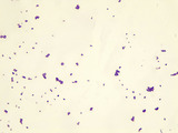
This micrograph was taken at 1000X total magnifcation on a brightfield microscope. The subject is Micrococcus cells grown on nutrient agar at 25 degrees Celsius. The cells were heat-fixed to a slide and Gram stained prior to visualization.Image credit: Emily Fox
This micrograph was taken at 1000X total magnifcation on a brightfield microscope. …
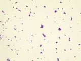
This micrograph was taken at 1000X total magnifcation on a brightfield microscope. The subject is Micrococcus luteus cells grown on agar at 37 degrees Celsius. The cells were heat-fixed to a slide and Gram stained prior to visualization.Image credit: Emily Fox
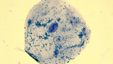
This micrograph was taken at 1000X total magnifcation on a brightfield microscope. …
This micrograph was taken at 1000X total magnifcation on a brightfield microscope. The subject is Micrococcus luteus cells grown on nutrient agar plates at 37 degrees Celsius. The cells were stained in a smear of nigrosin negative stain prior to visualization.Image credit: Emily Fox
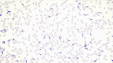
This micrograph was taken at 1000X total magnifcation on a brightfield microscope. …
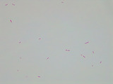
This micrograph was taken at 1000X total magnifcation on a brightfield microscope. The subject is Neisseria sicca cells grown on nutrient agar at 37 degrees Celsius. The cells were heat-fixed to a slide and Gram stained prior to visualization.Image credit: Emily Fox
This micrograph was taken at 1000X total magnifcation on a brightfield microscope. …
This micrograph was taken at 1000X total magnifcation on a brightfield microscope. The subject is Pseudomonas putida cells grown in broth culture overnight at 37 degrees Celsius. The cells were heat-fixed to a slide and Gram stained prior to visualization.Image credit: Emily Fox
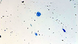
This micrograph was taken at 1000X total magnifcation on a brightfield microscope. …
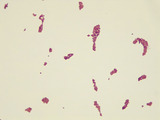
This micrograph was taken at 1000X total magnifcation on a brightfield microscope. The subject is Staphylococcus aureus cells grown on nutrient agar at 37 degrees Celsius. The cells were heat-fixed to a slide and Gram stained prior to visualization.Image credit: Emily Fox
This micrograph was taken at 1000X total magnifcation on a brightfield microscope. …
This micrograph was taken at 1000X total magnifcation on a brightfield microscope. The subject is Staphylococcus aureus cells grown on nutrient agar at 37 degrees Celsius. The cells were heat-fixed to a slide and Gram stained prior to visualization.Image credit: Emily Fox
This micrograph was taken at 400X total magnifcation on a brightfield microscope. …
This micrograph was taken at 400X total magnifcation on a brightfield microscope. The subject is giemsa-stained Trypanosoma cruzi in a blood smear.Image credit: Emily Fox
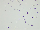
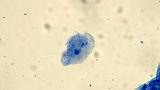
This micrograph was taken at 1000X total magnifcation on a brightfield microscope. …
This micrograph was taken at 1000X total magnifcation on a brightfield microscope. The subject is human cheek epithelial cells collected fresh with a toothpick. The cells were stained with methylene blue stain prior to visualization.Image credit: Emily Fox
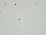
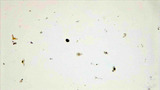
This micrograph was taken at 100X total magnifcation on a brightfield microscope. …
This micrograph was taken at 100X total magnifcation on a brightfield microscope. The subject is human cheek epithelial cells collected fresh with a toothpick. The cells were stained with methylene blue stain prior to visualization.Image credit: Emily Fox
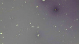
This micrograph was taken at 400X total magnifcation on a brightfield microscope. …
This micrograph was taken at 400X total magnifcation on a brightfield microscope. The subject is human cheek epithelial cells collected fresh with a toothpick. The cells were stained with methylene blue stain prior to visualization.Image credit: Emily Fox
With a simple list of necessary supplies, science teacher Mrs. Seay gets …
With a simple list of necessary supplies, science teacher Mrs. Seay gets her class completely involved in the task of identifying and classifying organisms found in local pond water.
No restrictions on your remixing, redistributing, or making derivative works. Give credit to the author, as required.
Your remixing, redistributing, or making derivatives works comes with some restrictions, including how it is shared.
Your redistributing comes with some restrictions. Do not remix or make derivative works.
Most restrictive license type. Prohibits most uses, sharing, and any changes.
Copyrighted materials, available under Fair Use and the TEACH Act for US-based educators, or other custom arrangements. Go to the resource provider to see their individual restrictions.