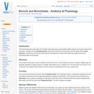
The trachea bifurcates at the levels of the 4th-6th intercostal space, approximately …
The trachea bifurcates at the levels of the 4th-6th intercostal space, approximately halfway between the thoracic inlet and the diaphragm. It divides into two principle bronchi, tubes which conduct air into the lungs, and they divide into two lobar bronchi for the left lung, and into four lobar bronchi for the right lung. These further divide into smaller bronchi and bronchioles within the lung tissue.
The Bursa of Fabricus is a primary lymphoid organ found in birds. …
The Bursa of Fabricus is a primary lymphoid organ found in birds. The bursa was the first place that a certain subset of lymphocytes was observed and consequently they were named B lymphocytes (bursa of Fabricius or bursa equivalent organs). The bursa is involved in the differentiation of B lymphocytes.
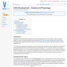
Development of the Central Nervous System (CNS) includes development of the brain, …
Development of the Central Nervous System (CNS) includes development of the brain, spinal cord, optic and auditory systems, as well as surrounding supporting cells including ependymal cells, astrocytes, oligodendrocytes and microglia. Information within this page will exclude development of the Peripheral Nervous System (PNS) which includes nerve and ganglia formation.
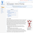
Blood is supplied to the brain from a ventral arterial supply in …
Blood is supplied to the brain from a ventral arterial supply in all species; from a circle of arteries called the Circle of Willis (also called the cerebral arterial circle or arterial circle of Willis) which lies ventrally to the hypothalamus where it forms a loose ring around the infundibular stalk. Although the appearance of the circle of Willis is fairly constant amongst mammals, the sources of blood supply to the circle and the direction of flow around the circle are very species specific. Blood is supplied to the brain by the internal carotid artery in dogs and horses whilst in other domestic species the main blood supply is from branches of the maxillary artery.
The cecum is a blind ending diverticulum of the large intestine and …
The cecum is a blind ending diverticulum of the large intestine and it exists at the junction of the ileum and the ascending colon. Its size and physiological importance varies between species. It is a site of microbial fermentation, absorption and transportation.
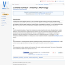
Camelids have a similar digestive structure to other ruminants, although camelids only …
Camelids have a similar digestive structure to other ruminants, although camelids only have three separate stomach compartments compared to the four found in domestic species. The first element of the camelid GI tract, known as C1, can be compared to the rumen whilst the second, known as C2 can be compared to the reticulum. The final element of the tract, C3 can be compared to the abomasum. Therefore camelids do not have a structure comparable to an omasum.
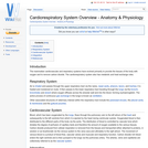
The mammalian cardiovascular and respiratory systems have evolved primarily to provide the …
The mammalian cardiovascular and respiratory systems have evolved primarily to provide the tissues of the body with oxygen and to remove carbon dioxide. The cardiorespiratory system also has metabolic and heat exchange roles.
Each mammary complex consists of 5-20 mammary units and their corresponding ducts. …
Each mammary complex consists of 5-20 mammary units and their corresponding ducts. The ducts open separately on the tip of the teat. Shallow grooves indicate the border between complexes. An intermammary sulcus divides the right from the left row.
The lower urinary tract is the collection of organs which convey the …
The lower urinary tract is the collection of organs which convey the formed urine from the kidneys to the exterior of the body. The urine is not altered in this part of the system in species other than the horse (where mucous is added) but instead its function is to collect and store the urine until enough of it is collected for release to become necessary. This gives the animal urinary continence. Three major structures make up this tract. The ureters, the bladder and the urethra.
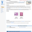
Also referred to as the reticuloendothelial system or mononuclear phagocytic system. It …
Also referred to as the reticuloendothelial system or mononuclear phagocytic system. It is comprised of primary lymphoid organs (bone marrow, Bursa of Fabricius, the foetal liver and the thymus) which are responsible for the production of lymphocytes, and the secondary lymphoid organs (lymph nodes, spleen and mucosal associated lymphoid tissue) which function to provide an environment where lymphocytes can react to antigen from the tissue fluid, blood and mucosal surfaces.
The musculoskeletal system includes bones, joints, cartilage, muscles, ligaments and tendons. In …
The musculoskeletal system includes bones, joints, cartilage, muscles, ligaments and tendons. In order to describe anatomical landmarks for example for the purposes of surgery and to be able to describe different directional information, for example when recording the view of a recently taken x-ray, it is necessary to have a way of describing the planes and axes that can be applied to the musculoskeletal system to pinpoint a specific anatomical area.
This page has links to information about pregnancy and parturation; including sperm …
This page has links to information about pregnancy and parturation; including sperm in the female tract, fertilisation, sexual differentiation, genital development, gestation lengths in different species, maternal recognition of pregnancy, litter sizes, placenta and its endocrine function, fetal circulation, puerperium, and reproductive disorders.
The Central Nervous System (CNS) is composed of the brain and the …
The Central Nervous System (CNS) is composed of the brain and the spinal cord. This page is specifically focussed on the histologic appearance, for anatomy see Forebrain, Midbrain, Hindbrain, Cranial Nerves, Spinal Cord and CNS Development.
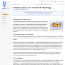
Cerebrospinal fluid (CSF) surrounds the brain as well as the central canal …
Cerebrospinal fluid (CSF) surrounds the brain as well as the central canal of the spinal cord. It helps cushion the central nervous system (CNS), acting in a similar manner to a shock absorber. It also acts as a chemical buffer providing immunological protection and a transport system for waste products and nutrients. The CSF also provides buoyancy to the soft neural tissues which effectively allows the neural tissue to "float" in the CSF. This prevents the brain tissue from becoming deformed under its own weight. It acts as a diffusion medium for the transport of neurotransmitters and neuroendocrine substances.
The cervix can be palpated transrectally and forms a sphincter controlling access …
The cervix can be palpated transrectally and forms a sphincter controlling access to the uterus.The anatomy of the cervical canal is adapted to suit a particular pattern of reproduction and its composition will alter under the influence of reproductive hormones. Not only does it respond to the fluctuation in oestrodiol during the oestrous cycle, but is responsive to prostaglandins and oxytocin in order to 'soften' for parturition.
The colon is a site of microbial fermentation, the relative importance of …
The colon is a site of microbial fermentation, the relative importance of this is species dependent. The colon can be divided into the following portions; Ascending, transverse and descending.
Different hormones, neurotransmitters and reflexes are involved in the complicated process of …
Different hormones, neurotransmitters and reflexes are involved in the complicated process of feeding in animals. Secretions and motility of the gastrointestinal tract are stimulated and carefully regulated by numerous factors, including environmental stimuli and the presence of food in different parts of the gastrointestinal tract from the oral cavity right through to the intestines. When a harmful substance is ingested the body acts to eliminate it in different ways to prevent the animal becoming ill, for example, through vomiting and diarrhoea. If one or more of the pathways in controlling feeding is damaged or inhibited, then problems such as obesity occurs.
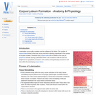
Corpus Luteum is latin for "yellow body". The corpus luteum is the …
Corpus Luteum is latin for "yellow body". The corpus luteum is the structure formed during luteinisation of the follicle after ovulation. The corpus luteum is, however, actually only yellow in the cow and in all other domestic species it is red. The yellow colouration of the corpus luteum is due to the pigment, lutein.
Luteinisation occurs after ovulation and the collapse of the follicle. The number …
Luteinisation occurs after ovulation and the collapse of the follicle. The number of corpora lutea formed in the ovary at any one time is directly proportional to the number of oocytes ovulated. Therefore many corpora lutea will be visible on the ovary of polytocous animals. During Luteinisation there is an increase in both the size and weight due to hyperplasia (increase in cell number) and hypertrophy (increase in cell size) within the developing corpus luteum.
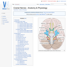
Cranial nerves are those nerves which arise from the brain and brain …
Cranial nerves are those nerves which arise from the brain and brain stem rather than the spinal cord. Nerves arising from the spinal cord are the peripheral nerves. There are 12 pairs of cranial nerves and these pairs of nerves passage through foramina in the skull, either individually or in groups. Cranial nerves are traditionally referred to by Roman numerals and these numerals begin cranially and run caudally.
No restrictions on your remixing, redistributing, or making derivative works. Give credit to the author, as required.
Your remixing, redistributing, or making derivatives works comes with some restrictions, including how it is shared.
Your redistributing comes with some restrictions. Do not remix or make derivative works.
Most restrictive license type. Prohibits most uses, sharing, and any changes.
Copyrighted materials, available under Fair Use and the TEACH Act for US-based educators, or other custom arrangements. Go to the resource provider to see their individual restrictions.