The vomeronasal organ is also known as the olfactory organ, or the …
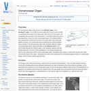
The vomeronasal organ is also known as the olfactory organ, or the Jacobson's organ. It is an olfactory sense organ that is found in most animals. It is positioned at the base of the nasal cavity, within the roof of the mouth, and is separated into two parts by the nasal septum. It is situated close to the vomer and nasal bones, hence the name vomeronasal organ.
The Guttural Pouch is present only in members of the order Perissodactyla …
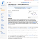
The Guttural Pouch is present only in members of the order Perissodactyla (nonruminant ungulates: horses, tapirs, rhinoceros) and another small band of small mammals including Hyraxes, certain bats and a South American mouse.
The trachea bifurcates at the levels of the 4th-6th intercostal space, approximately …
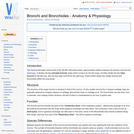
The trachea bifurcates at the levels of the 4th-6th intercostal space, approximately halfway between the thoracic inlet and the diaphragm. It divides into two principle bronchi, tubes which conduct air into the lungs, and they divide into two lobar bronchi for the left lung, and into four lobar bronchi for the right lung. These further divide into smaller bronchi and bronchioles within the lung tissue.
The surface of the inner wall of all of the body cavities …
The surface of the inner wall of all of the body cavities is lined by a serous membrane which consists of a single layer of flat epithelium with a thin underlying propria (connective tissue). Within the thoracic cavity, this is known as the pleura. The visceral pleura which coats the outer surface of the lung is derived from the splanchnic mesoderm. The parietal pleura lining the thoracic cavity is derived from somatic mesoderm. The pleural cavity is a potential space between the two areas of pleural membrane, which normally are adhesed to each other.
The air in the alveoli is renewed regularly, thanks to the ventilation …
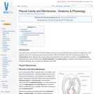
The air in the alveoli is renewed regularly, thanks to the ventilation process. Gas exchange in the lungs takes place between the blood in the capillary network surrounding the alveoli, and the air in the alveoli itself.
Nerves allow electrical impulses to propagate along their elongated cell extensions and …
Nerves allow electrical impulses to propagate along their elongated cell extensions and facilitate the transfer of information throughout the body. Neural tissue is found within the central nervous system (CNS) and the peripheral nervous system (PNS) and the composition and constituent parts of neurones and their surrounding cells differ only slightly.
Muscle cells can come from two lineages in the somite. Limb and …
Muscle cells can come from two lineages in the somite. Limb and body muscle develop from hypaxial muscle in the lateral regions of the somite. Back muscle develops from epaxial muscle in the dorsal regions of the somite. Muscle fibres have hundreds of nuclei and function as a syncytium.
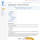
The adrenal glands are paired bodies lying cranial to the kidneys within …
The adrenal glands are paired bodies lying cranial to the kidneys within the retroperitoneal space. The glands consist of two layers; the cortex and medulla.
The adrenal glands are paired bodies lying cranial to the kidneys within …
The adrenal glands are paired bodies lying cranial to the kidneys within the retroperitoneal space. The glands consist of two layers; the cortex and medulla.
The integumentary system is an organ system that forms the protective covering …
The integumentary system is an organ system that forms the protective covering of an animal and comprises the skin (including glands and their products), haircoat or feathers, scales, nails, hooves and horns. The integumentary system has a variety of functions; in animals, it serves to waterproof, cushion and protect the deeper tissues, excrete waste, regulate temperature and is the location of sensory receptors for pain, pressure and temperature. Generally mammalian skin is covered with hair and is termed hirsute skin. Where hair is absent, it is termed glabrous skin.
The keratin in the epidermis, when cornified and thickened, is referred to …
The keratin in the epidermis, when cornified and thickened, is referred to as horn. Horn is particulary resistant to mechanical and chemical damage. The dermis of horn gives the structures their 3-D structure and shape. Cattle, some sheep, goats and antelope posess horns and these are permanent organs. Breeds without horns are termed polled breeds. Deer posess antlers, which are temporary organs that develop during the rutting season and are then shed.
The musculoskeletal system includes bones, joints, cartilage, muscles, ligaments and tendons. In …
The musculoskeletal system includes bones, joints, cartilage, muscles, ligaments and tendons. In order to describe anatomical landmarks for example for the purposes of surgery and to be able to describe different directional information, for example when recording the view of a recently taken x-ray, it is necessary to have a way of describing the planes and axes that can be applied to the musculoskeletal system to pinpoint a specific anatomical area.
The Central Nervous System (CNS) is composed of the brain and the …
The Central Nervous System (CNS) is composed of the brain and the spinal cord. This page is specifically focussed on the histologic appearance, for anatomy see Forebrain, Midbrain, Hindbrain, Cranial Nerves, Spinal Cord and CNS Development.
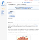
Cranial nerves are those nerves which arise from the brain and brain …
Cranial nerves are those nerves which arise from the brain and brain stem rather than the spinal cord. Nerves arising from the spinal cord are the peripheral nerves. There are 12 pairs of cranial nerves and these pairs of nerves passage through foramina in the skull, either individually or in groups. Cranial nerves are traditionally referred to by Roman numerals and these numerals begin cranially and run caudally.
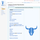
Reproduction is the complex set of biological processes that result in the …
Reproduction is the complex set of biological processes that result in the formation of a new organism; it is crucial that we understand how these processes occur normally and have a good grasp of the role of hormones in the reproductive process. Pathology and disease are common within the reproductive system and can not only lead to a sick animal but also to a loss in production. In this section we hope to cover all the main processes in domestic animals, laying a foundation for understanding the associated pathological states.
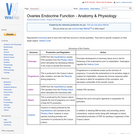
The ovary is the female Gonad homologous to the male Testes. It …
The ovary is the female Gonad homologous to the male Testes. It is usually a paired organ in domestic species, but in the bird only the left Ovary is present. The structures found within the ovary are undergoing constant changes throughout the oestrus cycle from the Follicles containing Oocytes, to the formation of Corpus Haemorrhagicum, Corpus Luteum, and finally Corpus Albicans. Ovaries are ellipsoidal in shape with an irregular surface due to the projection of dominant follicles and corpora lutea. These irregularities are absent in the mare due to the cortex and medulla being reversed with ovulation only occuring from the ovulation fossa. They are greatest in Polytocious animals such as the sow due to many dominant follicles, and so corpora lutea, developing at once.
Each mammary complex consists of 5-20 mammary units and their corresponding ducts. …
Each mammary complex consists of 5-20 mammary units and their corresponding ducts. The ducts open separately on the tip of the teat. Shallow grooves indicate the border between complexes. An intermammary sulcus divides the right from the left row.
Active transport is reliant on carrier proteins and thus follows the same …
Active transport is reliant on carrier proteins and thus follows the same rules as facilitated diffusion in that they are specific have a maximum rate and are subject to competition. Crucially they transport substances against their concentration gradient and so require energy to work.
No restrictions on your remixing, redistributing, or making derivative works. Give credit to the author, as required.
Your remixing, redistributing, or making derivatives works comes with some restrictions, including how it is shared.
Your redistributing comes with some restrictions. Do not remix or make derivative works.
Most restrictive license type. Prohibits most uses, sharing, and any changes.
Copyrighted materials, available under Fair Use and the TEACH Act for US-based educators, or other custom arrangements. Go to the resource provider to see their individual restrictions.