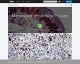
BCC Bioscience Image Library is a media file repository of images and …
BCC Bioscience Image Library is a media file repository of images and video clips made available to educators and students in the biological sciences. The resources are created by faculty, staff and students of Berkshire Community College and are licensed under Creative Commons 0. This means all content is free, with no restrictions on how the material may be used, reused, adapted or modified for any purposes, without restriction under copyright or database law.
This project was partially funded by a $20,000,000 grant awarded by the U.S. Department of Labor’s Employment and Training Administration, Grant # TC-26450-14-60-A-25. The product was created by the grantee and does not necessarily reflect the official position of the U.S. Department of Labor. The U.S. Department of Labor makes no guarantees, warranties, or assurances of any kind, express or implied, with respect to such information, including any information on linked sites and including, but not limited to, accuracy of the information or its completeness, timeliness, usefulness, adequacy, continued availability, or ownership.
If you have any questions contact professor Faye Reynolds at: freynold@berkshirecc.edu
Oogenesis is the process of producing the female gametes, the Ovum, from …
Oogenesis is the process of producing the female gametes, the Ovum, from the primordial germ cells. The majority of the steps in oogenesis, up to the point of producing primary oocytes, occur pre-natally. Therefore, females are born with all of the Primary Oocytes that they will ever have as primary oocytes do not divide further.
Camelids have a similar digestive structure to other ruminants, although camelids only …
Camelids have a similar digestive structure to other ruminants, although camelids only have three separate stomach compartments compared to the four found in domestic species. The first element of the camelid GI tract, known as C1, can be compared to the rumen whilst the second, known as C2 can be compared to the reticulum. The final element of the tract, C3 can be compared to the abomasum. Therefore camelids do not have a structure comparable to an omasum.
The duodenum is the proximal part of the small intestine and extends …
The duodenum is the proximal part of the small intestine and extends from the pylorus of the stomach to the jejunum. It has descending and ascending portions and both portions have digestive and absorptive functions.
The jejunum continues from the duodenum and leads into the ileum. It …
The jejunum continues from the duodenum and leads into the ileum. It is the longest part of the small intestine and is highly coiled. It has digestive and absorptive functions.
Deglutition is the process of swallowing. Food is passed from the oral …
Deglutition is the process of swallowing. Food is passed from the oral pharynx into the oesophageal/laryngeal pharynx whilst the epiglottis closes across the entrance of the trachea. It is an involuntary reflex preventing food from passing into the trachea and thus preventing choking and respiratory pneumonia.
Mastication is the process whereby food is broken down by mechanical digestion …
Mastication is the process whereby food is broken down by mechanical digestion in the oral cavity. The cheeks and tongue function to position food over the teeth, where grinding can occur. Mastication requires correct muscle movements and jaw articulation.
Blood vessel formation is a combination of the following three processes: Vasculogenesis: …
Blood vessel formation is a combination of the following three processes: Vasculogenesis: the formation of blood vessels from endothelial progenitor cells; Angiogenesis: the sprouting of new capillaries from pre-existing vessels; and Arteriogenesis: the remodelling of newly formed or pre-existing vascular channels into larger and more muscular arterioles.
Prior to birth the foetus is not capable of respiratory function and …
Prior to birth the foetus is not capable of respiratory function and thus relies on the maternal circulation to carry out gas, nutrient and waste exchange. The foetal and maternal blood never mix, instead they interface at the placenta. Consequently the liver and the lungs are non-functional, and a series of shunts exist in the foetal circulation so that these organs are almost completely by-passed.
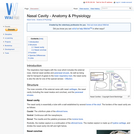
The respiratory tract begins with the nose which includes the external nose, …
The respiratory tract begins with the nose which includes the external nose, internal nasal cavities and paranasal sinuses. As well as being vital for transport of gases to the lower respiratory tract, the nasal cavity is also the site for one of the special senses - Olfaction.
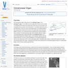
The vomeronasal organ is also known as the olfactory organ, or the …
The vomeronasal organ is also known as the olfactory organ, or the Jacobson's organ. It is an olfactory sense organ that is found in most animals. It is positioned at the base of the nasal cavity, within the roof of the mouth, and is separated into two parts by the nasal septum. It is situated close to the vomer and nasal bones, hence the name vomeronasal organ.
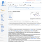
The Guttural Pouch is present only in members of the order Perissodactyla …
The Guttural Pouch is present only in members of the order Perissodactyla (nonruminant ungulates: horses, tapirs, rhinoceros) and another small band of small mammals including Hyraxes, certain bats and a South American mouse.
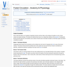
The trachea bifurcates at the levels of the 4th-6th intercostal space, approximately …
The trachea bifurcates at the levels of the 4th-6th intercostal space, approximately halfway between the thoracic inlet and the diaphragm. It divides into two principle bronchi, tubes which conduct air into the lungs, and they divide into two lobar bronchi for the left lung, and into four lobar bronchi for the right lung. These further divide into smaller bronchi and bronchioles within the lung tissue.
The air in the alveoli is renewed regularly, thanks to the ventilation …
The air in the alveoli is renewed regularly, thanks to the ventilation process. Gas exchange in the lungs takes place between the blood in the capillary network surrounding the alveoli, and the air in the alveoli itself.
Muscle cells can come from two lineages in the somite. Limb and …
Muscle cells can come from two lineages in the somite. Limb and body muscle develop from hypaxial muscle in the lateral regions of the somite. Back muscle develops from epaxial muscle in the dorsal regions of the somite. Muscle fibres have hundreds of nuclei and function as a syncytium.
Teeth develop differently in different regions of the mouth in most species, …
Teeth develop differently in different regions of the mouth in most species, a process called heterodonty. In some animals, teeth develop identically in different regions of the mouth, a process called homodonty. Different species will have varying numbers of teeth and different shapes depending largely on their diet. Not all species possess teeth and there is huge variation in dental formulae between the species that have teeth. Teeth are mainly used for mastication - chewing and grinding food particles, but are also used for seizing prey and tearing. The occlusion surface is where opposing teeth touch. The contact surface is where adjacent teeth touch.
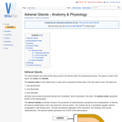
The adrenal glands are paired bodies lying cranial to the kidneys within …
The adrenal glands are paired bodies lying cranial to the kidneys within the retroperitoneal space. The glands consist of two layers; the cortex and medulla.
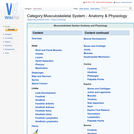
The musculoskeletal system includes bones, joints, cartilage, muscles, ligaments and tendons. In …
The musculoskeletal system includes bones, joints, cartilage, muscles, ligaments and tendons. In order to describe anatomical landmarks for example for the purposes of surgery and to be able to describe different directional information, for example when recording the view of a recently taken x-ray, it is necessary to have a way of describing the planes and axes that can be applied to the musculoskeletal system to pinpoint a specific anatomical area.
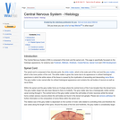
The Central Nervous System (CNS) is composed of the brain and the …
The Central Nervous System (CNS) is composed of the brain and the spinal cord. This page is specifically focussed on the histologic appearance, for anatomy see Forebrain, Midbrain, Hindbrain, Cranial Nerves, Spinal Cord and CNS Development.
No restrictions on your remixing, redistributing, or making derivative works. Give credit to the author, as required.
Your remixing, redistributing, or making derivatives works comes with some restrictions, including how it is shared.
Your redistributing comes with some restrictions. Do not remix or make derivative works.
Most restrictive license type. Prohibits most uses, sharing, and any changes.
Copyrighted materials, available under Fair Use and the TEACH Act for US-based educators, or other custom arrangements. Go to the resource provider to see their individual restrictions.