Updating search results...

Search Resources

11 Results

View
Selected filters:
  • actin
AKT2 deficiency impairs formation of the BCR
signalosome
Unrestricted Use
CC BY
Rating
0.0 stars

This resource is a video abstract of a research paper created by Research Square on behalf of its authors. It provides a synopsis that's easy to understand, and can be used to introduce the topics it covers to students, researchers, and the general public. The video's transcript is also provided in full, with a portion provided below for preview:

"Some proteins are central to many cell signaling processes. One of these key molecules is AKT2. An important kinase involved in cell survival, growth, and metabolism, it has ties to insulin-induced signaling and cancer. AKT2 has a critical role in immune cells such as neutrophils and macrophages; however, although AKT2 is expressed in antibody-producing immune cells called B cells, its function in B cells isn’t clear. In a recent study, researchers sought to understand the role of AKT2 in B cells using AKT2-deficient mice. They found that mice lacking AKT2 had impaired B-cell differentiation. B cells from these mice were not able to form a cluster of molecules called a signalosome in response to B-cell receptor (BCR) signaling, resulting in poor BCR signaling and impaired B cell activation and spreading. These results suggest that as a central orchestrator of signaling, AKT2 function is critical for proper BCR signaling and B cell development, ensuring a functional antibody-mediated immune response..."

The rest of the transcript, along with a link to the research itself, is available on the resource itself.

Subject:
Biology
Life Science
Material Type:
Diagram/Illustration
Reading
Provider:
Research Square
Provider Set:
Video Bytes
Date Added:
06/23/2020
Abl kinase’s disordered region plays roles in protein function and stability
Unrestricted Use
CC BY
Rating
0.0 stars

This resource is a video abstract of a research paper created by Research Square on behalf of its authors. It provides a synopsis that's easy to understand, and can be used to introduce the topics it covers to students, researchers, and the general public. The video's transcript is also provided in full, with a portion provided below for preview:

"The non-receptor tyrosine kinase Abelson (Abl) is a key player in oncogenesis, causing diseases including chronic myelogenous and acute lymphoblastic leukemia. Drugs targeting Abl kinase activity serve as paradigms of targeted therapy. Drosophila is an ideal model for studying Abl’s function because there is only a single fly Abl family member. In flies, Abl is essential for embryonic morphogenesis, playing diverse roles in embryonic and adult viability. To examine the role of the intrinsically disordered region (IDR) of Abl, researchers deleted the IDR in Drosophila. They found that Abl lacking the IDR was not able to rescue the roles of Abl in viability and embryonic morphogenesis. The IDR was also essential for cell shape changes and cytoskeletal regulation during embryonic morphogenesis and, surprisingly, for modulating protein stability..."

The rest of the transcript, along with a link to the research itself, is available on the resource itself.

Subject:
Biology
Life Science
Material Type:
Diagram/Illustration
Reading
Provider:
Research Square
Provider Set:
Video Bytes
Date Added:
10/16/2021
Immune Cell Migration: On the Move in Response to Pathogens and Cancer Immunotherapy
Conditional Remix & Share Permitted
CC BY-NC-SA
Rating
0.0 stars

The mammalian immune system is sometimes called a “liquid organ,” capable of rapidly initiating and then resolving potent responses to pathogens at almost any location in the organism. What protein machinery drives immune cells’ rapid migration? How do cells make pathfinding decisions around barriers? How do they find rare pathogens or target cells in complex environments?
This course will begin by examining the general immunological functions of two major immune cell types—T cells and dendritic cells. Through our readings and discussions, we will examine the connections between immunotherapy as an emerging treatment modality for a variety of cancers and the migration of immune cells.
This course is one of many Advanced Undergraduate Seminars offered by the Biology Department at MIT. These seminars are tailored for students with an interest in using primary research literature to discuss and learn about current biological research in a highly interactive setting. Many instructors of the Advanced Undergraduate Seminars are postdoctoral scientists with a strong interest in teaching.

Subject:
Applied Science
Biology
Health, Medicine and Nursing
Life Science
Physical Science
Material Type:
Full Course
Provider:
MIT
Provider Set:
MIT OpenCourseWare
Author:
Fessenden, Timothy
Date Added:
09/01/2021
MACC1 driven alterations in cellular biomechanics facilitate cell motility in glioblastoma
Unrestricted Use
CC BY
Rating
0.0 stars

This resource is a video abstract of a research paper created by Research Square on behalf of its authors. It provides a synopsis that's easy to understand, and can be used to introduce the topics it covers to students, researchers, and the general public. The video's transcript is also provided in full, with a portion provided below for preview:

"Glioblastoma (GBM), an aggressive cancer in the brain or spinal cord, is a devastating diagnosis. Although therapies exist, GBM has a poor prognosis, with a median survival of only 14-15 months after diagnosis. Key to its aggressiveness is the degree to which migrating GBM cells infiltrate adjacent brain tissue. GBM cells express the protein MACC1, which is a marker of metastasis and tumor cell migration. Unfortunately, how GBM cells learn to migrate is unclear. A recent study used live-cell and atomic force microscopy to evaluate cell migration and mechanical properties of GBM cells overexpressing MACC1. The results showed that MACC1 increased the migratory speed and elasticity of GBM cells while it decreased cell-cell adhesion and inhibited aggregation. MACC1-overexpressing cells also had specific increases in protrusive actin, allowing the cells to adhere to laminin..."

The rest of the transcript, along with a link to the research itself, is available on the resource itself.

Subject:
Biology
Life Science
Material Type:
Diagram/Illustration
Reading
Provider:
Research Square
Provider Set:
Video Bytes
Date Added:
10/30/2020
Neuroprotective functions of the HDAC6 zinc finger ubiquitin-binding domain
Unrestricted Use
CC BY
Rating
0.0 stars

This resource is a video abstract of a research paper created by Research Square on behalf of its authors. It provides a synopsis that's easy to understand, and can be used to introduce the topics it covers to students, researchers, and the general public. The video's transcript is also provided in full, with a portion provided below for preview:

"Alzheimer’s disease and similar neurodegenerative diseases involve aggregation of the protein Tau and disruption of cell structural networks. The protein HDAC6 helps clear Tau aggregates and regulate the cytoskeleton, thus exerting neuroprotective effects. HDAC6’s catalytic domains mediate some of these functions, but the roles of another domain, the zinc finger ubiquitin-binding domain (ZnF UBP), are less understood. A recent study investigated the effects of purified HDAC6 ZnF UBP on cultured neuronal cells. The researchers found that HDAC6 ZnF UBP was nontoxic to cells, and cell imaging showed that it promoted reorganization of the cytoskeletal components actin and tubulin in ways that likely support neuron growth and migration. Localization of the protein ApoE in cell nuclei was increased, indicating improved neuronal health..."

The rest of the transcript, along with a link to the research itself, is available on the resource itself.

Subject:
Biology
Life Science
Material Type:
Diagram/Illustration
Reading
Provider:
Research Square
Provider Set:
Video Bytes
Date Added:
10/14/2021
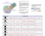
The Online Macromolecular Museum
Rating
0.0 stars

The Online Macromolecular Museum (OMM) is a site for the display and study of macromolecules. Macromolecular structures, as discovered by crystallographic or NMR methods, are scientific objects in much the same sense as fossil bones or dried specimens: they can be archived, studied, and displayed in aesthetically pleasing, educational exhibits. Hence, a museum seems an appropriate designation for the collection of displays that we are assembling. The OMM's exhibits are interactive tutorials on individual molecules in which hypertextual explanations of important biochemical features are linked to illustrative renderings of the molecule at hand.

Why devote a site to detailed visualizations of different macromolecules? In learning about the intricacies of life processes at the molecular level, it is important to understand how natural selection has fashioned the structure and chemistry of macromolecular machines to suit them for particular functions. This understanding is greatly facilitated by the visualization of 3-dimensional structure, when known. So, if static views of molecules (even in stereo) are worth a thousand words, then interactive animations of molecules should be worth much more. Indeed, we have found the types of displays represented here invaluable in gaining an appreciation for the details of key biochemical processes.

As Carl Brandon and John Tooze stated in their classic text, Introduction to Protein Structure:
"Molecular biology began some 40 years ago with the realization that structure was crucial for a proper understanding of function. Paradoxically, the dazzling achievements of molecular genetics and biochemistry led to the eclipse of structural studies. We believe the wheel has now come full circle, and those very achievements have increased the need for structural analysis at the same time that they have provided the means for it."

It is our opinion that structural analysis should extend into the classroom: as students learn about cellular mechanisms it is important that they study the chemistry of the molecular machines involved. These considerations have motivated the construction of the OMM.

The OMM is part of a collaborative effort by faculty and students interested in macromolecular structure-function relationships. The primary authors of some tutorials are students of David Marcey and he serves as author, co-author and site editor, and assumes all responsibility for content. Any criticisms, suggestions, comments, or questions should be sent to him at: marcey@callutheran.edu. All tutorials are copyrighted.

The OMM was started in 1996 for a Molecular Biology class at Kenyon College, where DM was a professor in the Biology Department (1990-1999). The OMM is now developed and housed at California Lutheran University, where DM has been a professor since 1999.

Subject:
Chemistry
Life Science
Physical Science
Material Type:
Activity/Lab
Diagram/Illustration
Homework/Assignment
Interactive
Lesson
Author:
David Marcey
Date Added:
09/28/2017
PIM1 accelerates prostate cancer cell motility by phosphorylating actin capping proteins
Unrestricted Use
CC BY
Rating
0.0 stars

This resource is a video abstract of a research paper created by Research Square on behalf of its authors. It provides a synopsis that's easy to understand, and can be used to introduce the topics it covers to students, researchers, and the general public. The video's transcript is also provided in full, with a portion provided below for preview:

"Prostate cancer is one of the prevalent forms of cancer in men around the world. Prostate tumors become especially fatal when they spread to one and other vital organs. The main forces driving cell motility are the constant polymerization and depolymerization of actin filaments which lead to changes in cellular protrusions. This process is regulated by actin-binding proteins, such as capping proteins, which bind to the fast-growing ends of actin filaments. A new study shows that these capping proteins are substrates for cancer-inducing PIM kinases, proteins whose overexpression promotes cancer cell survival and motility. Phosphorylation of capping proteins decreases their ability to protect actin filament ends from disassembly, leading to enhanced cell motility. while PIM inhibitors or capping protein mutations have the opposite effect, reducing cell migration..."

The rest of the transcript, along with a link to the research itself, is available on the resource itself.

Subject:
Biology
Life Science
Material Type:
Diagram/Illustration
Reading
Provider:
Research Square
Provider Set:
Video Bytes
Date Added:
11/11/2020
Phosphoinositide Signaling Modulates Microglial Phagocytosis in Alzheimer’s disease
Unrestricted Use
CC BY
Rating
0.0 stars

This resource is a video abstract of a research paper created by Research Square on behalf of its authors. It provides a synopsis that's easy to understand, and can be used to introduce the topics it covers to students, researchers, and the general public. The video's transcript is also provided in full, with a portion provided below for preview:

"Alzheimer’s disease affects 1 in 3 senior citizens worldwide. The hallmark of Alzheimer’s is the accumulation of abnormal protein deposits, including Tau and amyloid β, in neurons and glial cells in the brain. These deposits disrupt signal transduction by affecting lipid-based secondary messengers in the brain called phosphatidylinositols (PIs). PIs drive the reorganization of the cytoskeleton in glial cells, affecting many cellular processes. These dynamic molecules are tightly regulated by their phosphorylation status, which influences their abundance and localization. Because microglia in the brain must respond to chemotactic and pro-inflammatory signals, disrupting PIs alters microglial function, resulting in hyperactivation and inflammation. PI signaling typically drives actin remodeling to modulate phagocytosis, allowing glial cells to clear amyloid β aggregates and debris. Unfortunately, extensive amyloid β accumulation disrupts PI signaling, altering cytoskeleton regulation..."

The rest of the transcript, along with a link to the research itself, is available on the resource itself.

Subject:
Biology
Life Science
Material Type:
Diagram/Illustration
Reading
Provider:
Research Square
Provider Set:
Video Bytes
Date Added:
10/16/2021
Roles of SRC family kinases in epithelial–mesenchymal transition
Unrestricted Use
CC BY
Rating
0.0 stars

This resource is a video abstract of a research paper created by Research Square on behalf of its authors. It provides a synopsis that's easy to understand, and can be used to introduce the topics it covers to students, researchers, and the general public. The video's transcript is also provided in full, with a portion provided below for preview:

"Since the first SRC family kinase (SFK) was discovered in 1911, numerous SFKs have been identified in humans. SFKs are expressed in diverse tissues and cell types, where they regulate various biological processes. Posttranslational modification of these membrane-associated kinases can regulate their activity to affect cell signaling in different ways, but dysregulation of SFKs can promote cancer progression, invasion, and metastasis. In cancer cells, the kinases can promote epithelial-to-mesenchymal transition (EMT) to promote invasion. Specifically, they destabilize cell junctions, change cell polarity, and mediate invadopodia formation by influencing the actin cytoskeleton. SFKs also activate EMT-inducing molecules by influencing signaling pathways such as the TGF-β/SMAD, Wnt, NOTCH, and EGFR pathways. Given the roles of SFKs in cancer, SFK inhibitors have been developed as therapies for metastatic disease..."

The rest of the transcript, along with a link to the research itself, is available on the resource itself.

Subject:
Biology
Life Science
Material Type:
Diagram/Illustration
Reading
Provider:
Research Square
Provider Set:
Video Bytes
Date Added:
10/13/2021
Using mechanobiology and materials methods in epithelial-mesenchymal transition research
Unrestricted Use
CC BY
Rating
0.0 stars

This resource is a video abstract of a research paper created by Research Square on behalf of its authors. It provides a synopsis that's easy to understand, and can be used to introduce the topics it covers to students, researchers, and the general public. The video's transcript is also provided in full, with a portion provided below for preview:

"Cancer-related mortality, a leading cause of death in the US, is driven by tumor invasion and metastasis. Implicated in these processes is epithelial-to-mesenchymal transition, or EMT. EMT drives invasion through a dramatic reorganization of a cell's cytoskeleton and the extracellular matrix. Because EMT is a rare event, undergone by a few abnormal cells, it is difficult to view directly in a patient. But new research methods are providing a lens into this critical process. Culturing cells on planar surfaces is revealing how their EMT behavior is coordinated and driven by leader cells. Research on the protein vimentin highlights its role in enabling cells to contort during migration or proliferation. Other studies examine how topographically patterning culture surfaces changes the behaviors of cells as they slip into and out of EMT. And 3D matrices are being used to examine the dissemination and disorganization of multicellular clusters..."

The rest of the transcript, along with a link to the research itself, is available on the resource itself.

Subject:
Biology
Life Science
Material Type:
Diagram/Illustration
Reading
Provider:
Research Square
Provider Set:
Video Bytes
Date Added:
03/11/2021